
XIENCE Sierra
Everolimus Eluting Coronary Stent System
en
English
de
Deutsch
sv
Svenska
da
Dansk
pl
Polski
cs
Česky
tr
Türkçe
no
Norsk
it
Italiano
fi
Suomi
fr
Français
es
Español
ru
Pусский
pt
Português
nl
Nederlands
el
Ελληνικά
hu
Magyar
bg
Български
ro
Română
sk
Slovensky
et
Eesti keel
lv
Latviešu
lt
Lietuvių
sl
Slovenščina
uk
Українська
id
Bahasa Indonesia
ar
Graphical Symbols for Medical Device Labeling; Grafische Symbole zur Beschriftung von Medizinprodukten; Symboles graphiques pour l’étiquetage des dispositifs
médicaux ; Símbolos gráficos utilizados en el etiquetado de productos sanitarios; Simboli grafici per l’etichettatura di dispositivi medicali; Símbolos Gráficos para
Rotulagem de Dispositivos Médicos; Grafiska symboler för märkning av medicinsk utrustning; Grafische symbolen voor de etikettering van medische hulpmiddelen;
Grafiske symboler beregnet til mærkning af medicinske produkter; Γραφικά σύμβολα για τη σήμανση ιατρικών συσκευών
Symbole graficzne do oznaczania sprzętu medycznego; Grafikus szimbólumok orvosi eszközök címkézéséhez; Grafické symboly k označení zdravotnického prostředku;
Tıbbi Cihaz Etiketleri için Grafik Semboller; Grafické symboly na označovanie zdravotníckej pomôcky
0086
Графични символи за етикетиране на медицински уреди; Simboluri grafice pentru etichetarea dispozitivelor medicale; Графические символы на этикетках
медицинских изделий; Lääketieteellisten laitteiden tuotetarroissa esiintyvät symbolit
Grafiske symboler for merking av medisinsk utstyr; Meditsiiniseadme märgistuse graafilised sümbolid; Grafiskie simboli medicīnisko ierīču apzīmēšanai; Medicinos
prietaisų etikečių grafiniai simboliai; Grafični simboli za označevanje medicinskega pripomočka
Графічні позначки на етикетках медичних виробів; Graphical Symbols for Medical Device Labeling;
EL2115538 (2017-11-03)
Page 1 of 206
Printed on : 2017-11-03

Catalogue number; Katalog-Nr.; N° de référence ; N.
o
de
referencia; N. di catalogo; Número de catálogo; Katalognr;
Catalogusnummer; Katalognummer; Aριθμός καταλόγου
Manufacturer; Hersteller; Fabricant ; Fabricante; Produttore;
Fabricante; Tillverkare; Fabrikant; Producent; Κατασκευαστής
French size; French-Größe; Taille en French ; Calibre French;
Calibro in French; Tamanho em French (F); French-storlek;
Maateenheid French; French størrelse; Μέγεθος σε French
Sterilized using ethylene oxide; Mit Ethylenoxid sterilisiert;
Stérilisé à l’oxyde d’éthylène ; Esterilizado con óxido de
etileno; Sterilizzato con ossido di etilene; Esterilizado por
óxido de etileno; Steriliserad med etylenoxid; Gesteriliseerd
met ethyleenoxide; Steriliseret med ethylenoxid;
Αποστειρωμένο με οξείδιο του αιθυλενίου
Consult instructions for use; Gebrauchsanweisung lesen;
Consulter le mode d’emploi ; Consultar las instrucciones
de uso; Consultare le istruzioni per l’uso; Consultar as
instruções de utilização; Se bruksanvisningen; Raadpleeg
gebruiksaanwijzing; Læs brugsanvisningen; Συμβουλευτείτε
τις οδηγίες χρήσης
Use by; Verwenden vor; Date limite ; Fecha de caducidad;
Data di scadenza; Usar até; Bäst före; Uiterste gebruiksdatum;
Anvendes inden; Χρήση έως
Batch code; Chargencode; Nº de lot ; Código de lote;
Codice Lotto; Código do lote; Batchnummer; Partijnummer;
Partinummer; Αριθμός παρτίδας
Date of manufacture; Herstellungsdatum; Date de fabrication ;
Fecha de fabricación; Data di produzione; Data de fabrico;
Tillverkningsdatum; Productiedatum; Fremstillingsdato;
Ημερομηνία κατασκευής
Graphical Symbols for Medical Device Labeling; Grafische Symbole zur Beschriftung von Medizinprodukten; Symboles graphiques pour l’étiquetage des dispositifs médicaux ; Símbolos
gráficos utilizados en el etiquetado de productos sanitarios; Simboli grafici per l’etichettatura di dispositivi medicali; Símbolos Gráficos para Rotulagem de Dispositivos Médicos;
Grafiska symboler för märkning av medicinsk utrustning; Grafische symbolen voor de etikettering van medische hulpmiddelen; Grafiske symboler beregnet til mærkning af medicinske
produkter; Γραφικά σύμβολα για τη σήμανση ιατρικών συσκευών
Authorised representative in the European Community;
Bevollmächtigte Vertretung in der Europäischen
Gemeinschaft; Représentant agréé pour la Communauté
européenne ; Representante autorizado en la Comunidad
Europea; Rappresentante autorizzato nella Comunità
europea; Representante autorizado na Comunidade Europeia;
Auktoriserad representant i Europeiska gemenskapen;
In de Europese Gemeenschap gevestigde gemachtigde;
Autoriseret repræsentant i Det Europæiske Fællesskab;
Εξουσιοδοτημένος αντιπρόσωπος στην Ευρωπαϊκή
Kοινότητα
Outer diameter; Außendurchmesser; Diamètre externe ;
Diámetro externo; Diametro esterno; Diâmetro Externo;
Ytterdiameter; Buitendiameter; Udvendig diameter;
Εξωτερική διάμετρος
Inner diameter; Innendurchmesser; Diamètre interne ;
Diámetro interno; Diametro interno; Diâmetro interno;
Innerdiameter; Binnendiameter; Indvendig diameter;
Εσωτερική διάμετρος
Stent length; Stentlänge; Longueur de l’endoprothèse ;
Longitud del stent; Lunghezza dello stent; Comprimento do
stent; Stentlängd; Stentlengte; Stentlængde; Μήκος του στεντ
EL2115538 (2017-11-03)
Page 2 of 206
Printed on : 2017-11-03

Do not resterilize; Nicht resterilisieren; Ne pas restériliser ;
No volver a esterilizar; Non risterilizzare; Não reesterilizar;
Får ej omsteriliseras; Niet opnieuw steriliseren; Må ikke
resteriliseres; Μην τo επαναποστειρώνετε
Do not reuse; Nicht wiederverwenden; Ne pas réutiliser ;
No volver a utilizar; Monouso; Não reutilizar; Får ej
återanvändas; Niet opnieuw gebruiken; Må ikke genbruges;
Μην το επαναχρησιμοποιείτε
Guiding catheter; Führungskatheter; Cathéter-guide ;
Catéter guía; Catetere guida; Cateter-Guia; Guidekateter;
Geleidekatheter; Guidingkateter; Οδηγός καθετήρας
Non-pyrogenic; Nicht pyrogen; Apyrogène ; Apirógeno;
Apirogeno; Apirogénico; Icke pyrogen; Niet-pyrogeen;
Non-pyrogen; Μη πυρετογόνο
Packaging unit; Packungseinheit; Unité de conditionnement ;
Unidad de envasado; Unità di imballaggio; Unidade de
embalagem; Förpackningsenhet; Verpakkingseenheid;
Pakkeenhed; Μονάδα συσκευασίας
MR Conditional; Bedingt MRT-kompatibel; Compatible avec
l’IRM sous conditions ; Compatibilidad condicionada con
la RM; A compatibilità RM condizionata; Condicionado
ao ambiente de ressonância magnética; MR-säker under
specifika betingelser; Onder bepaalde voorwaarden MR-veilig;
MR-betinget; Συμβατό με μαγνητική τομογραφία
MR Conditional
Do not use if package is damaged; Das Produkt nicht
verwenden, wenn die Packung beschädigt ist; Ne pas
utiliser ce produit si l'emballage est endommagé ; No utilizar
el producto si el envase está dañado; Non utilizzare se la
confezione è danneggiata; Não utilizar se a embalagem
estiver danificada; Använd inte produkten om förpackningen
är skadad; Gebruik het product niet als de verpakking
beschadigd is; Må ikke anvendes, hvis pakningen er
beskadiget; Μη χρησιμοποιείτε το προϊόν, εάν η συσκευασία
είναι κατεστραμμένη
Stent post-dilatation limit; Nachdilatationsgrenze des Stents;
Limite post-dilatation de l’endoprothèse ; Límite del stent tras
la dilatación; Limite di post-dilatazione dello stent; Limite
pós-dilatação do stent; Stentens postdilatationsgräns;
Postdilatatielimiet van stent; Grænse for postdilatation af
stenten; Όριο μεταδιαστολής στεντ
Temperature limitation; Temperaturbegrenzung; Limites
de température; Límites de temperatura; Limiti di
temperatura; Limites de temperatura; Temperaturgräns;
Temperatuurbegrenzing; Temperaturbegrænsning;
Περιορισμός θερμοκρασίας
25˚C
(77˚F)
Keep away from sunlight; Vor Sonnenlicht schützen;
Conserver à l’abri de la lumière du soleil ; No exponer a la luz
del sol; Tenere al riparo dalla luce del sole; Guardar ao abrigo
da luz solar; Får inte utsättas för solljus; Uit de buurt van
zonlicht houden; Beskyttes mod sollys; Διατηρείτε το μακριά
από το ηλιακό φως
Keep dry; Trocken halten; Conserver au sec ; Mantener seco;
Mantenere asciutto; Manter seco; Förvaras torrt; Droog
houden; Opbevares tørt; Διατηρείτε το στεγνό
Excursions permitted to temperature range; Toleranzbereich
der Temperatur; Expositions autorisées à la plage de
température ; Variaciones permitidas del intervalo de
temperatura; Escursioni termiche ammesse fino all'intervallo
di temperatura; Amplitude de temperatura permitida;
Tillfälliga avvikelser tillåtna inom temperaturområdet; Bereik
toegestane temperatuurschommelingen; Udsving tilladt for
temperaturinterval; Επιτρεπόμενες διακυμάνσεις εύρους
θερμοκρασίας
EL2115538 (2017-11-03)
Page 3 of 206
Printed on : 2017-11-03

English
8.0 PATIENT SELECTION AND TREATMENT
8.1 Individualization of Treatment
9.0 CLINICIAN USE INFORMATION
9.1 Inspection Prior to Use
9.2 Materials Required
9.3 Preparation
9.3.1 Packaging Removal
9.3.2 Guide Wire Lumen Flush
9.3.3 Delivery System Preparation
9.4 Delivery Procedure
9.5 Deployment Procedure
9.6 Removal Procedure
9.7 Post-deployment Dilation of Stent Segments
10.0 SPIRIT AND XIENCE FAMILY OF CLINICAL TRIALS
10.1 Pre-Market Clinical Trials
Table 10.1-1: SPIRIT Family of XIENCE V Clinical Trial Designs (Pre-market)
Table 10.1-2: SPIRIT Family of Clinical Trials Angiographic Results (Pre-market)
Table 10.1-3: SPIRIT Family of Clinical Trials Principal 1-Year Clinical Outcomes
(Pre-market)
Table 10.1-4: SPIRIT Family of Clinical Trials Principal Clinical Outcomes from
Latest Follow-up (Pre-market)
10.2 Post-Market Clinical Trials
Table 10.2-1: SPIRIT and XIENCE V Family Clinical Trial Designs (Post-market)
Table 10.2-2: SPIRIT and XIENCE V Family of Trials Principal Clinical Outcomes
(Post-market)
10.3 Pooled Clinical Trials Analysis for Stent Thrombosis Subsequent to DAPT
Interruption / Discontinuation
Table 10.3-1: Stent Thrombosis (ARC Definite / Probable) Subsequent to DAPT
Interruption / Discontinuation through 730 days Versus No Interruption; Pooled Data
from 7 Pre- and Post-market Trials
11.0 HOW SUPPLIED
12.0 PATENTS AND TRADEMARKS
XIENCE Sierra
Everolimus Eluting Coronary Stent System
INFORMATION FOR PRESCRIBERS
Table of Contents
1.0 DEVICE DESCRIPTION
Table 1-1: Product Name and Sizes
Table 1-2: Drug Content in the XIENCE Sierra
Everolimus Eluting Coronary Stents
Table 1-3: In vitro Device Specifications
2.0 INDICATIONS
3.0 CONTRAINDICATIONS
4.0 WARNINGS
5.0 PRECAUTIONS
5.1 Stent Handling
5.2 Stent Placement
5.3 Use in Conjunction with Other Procedures
5.4 Stent / System Removal
5.5 Post Implant
5.6 Use in Special Populations
5.6.1 Pregnancy
5.6.2 Lactation
5.6.3 Pediatric use
5.7 Magnetic Resonance Imaging Safety Information (MRI)
5.8 Drug Interactions
5.9 Immune Suppression Potential
5.10 Lipid Elevation Potential
6.0 DRUG INFORMATION
6.1 Interactions with Drugs or Other Substances
6.2 Pregnancy
6.3 Lactation
7.0 POTENTIAL ADVERSE EVENTS
• Two radiopaque markers, located underneath the balloon, fluoroscopically mark the working length of the balloon and the expanded stent length.
• Two proximal delivery system shaft markers (95 cm and 105 cm proximal to the distal tip) indicate the relative position of the delivery system to the end of the brachial or femoral
guiding catheter. Working catheter length is 145 cm.
• A shaft color change denotes the guide wire exit notch.
Table 1-3: In vitro Device Specifications
Stent Diameter
(mm)
Stent Length
(mm)
* Minimum
Guiding Catheter Compatibility (ID)
** in vitro Stent Nominal Pressure Rated Burst Pressure (RBP)
Stent Free
Area
(%)
(atm) kPa (atm) kPa
2.0 8, 12, 15, 18, 23, 28, 33, 38 5F (0.056” / 1.42 mm) 9 912 16 1621 81
2.25 8, 12, 15, 18, 23, 28, 33, 38 5F (0.056” / 1.42 mm) 9 912 16 1621 83
2.5 8, 12, 15, 18, 23, 28, 33, 38 5F (0.056” / 1.42 mm) 9 912 16 1621 84
2.75 8, 12, 15, 18, 23, 28, 33, 38 5F (0.056” / 1.42 mm) 12 1216 16 1621 86
3.0 8, 12, 15, 18, 23, 28, 33, 38 5F (0.056” / 1.42 mm) 12 1216 16 1621 87
3.25 8, 12, 15, 18, 23, 28, 33, 38 5F (0.056” / 1.42 mm) 12 1216 16 1621 88
3.5 8, 12, 15, 18, 23, 28, 33, 38 5F (0.056” / 1.42 mm) 12 1216 16 1621 84
4.0 8, 12, 15, 18, 23, 28, 33, 38 5F (0.056” / 1.42 mm) 12 1216 16 1621 86
* See individual manufacturer specifications for (F) equivalent.
** Assure full deployment of the stent (see Section 9.5 Deployment Procedure). Deployment pressures should be based on lesion characteristics.
2.0 INDICATIONS
The XIENCE Sierra Everolimus Eluting Coronary Stent System is indicated for improving coronary luminal diameter in the following:
• Patients with symptomatic ischemic heart disease due to discrete de novo native coronary artery lesions.
• For restoring coronary flow in patients experiencing acute myocardial infarction who present within 12 hours of symptom onset.
• For the treatment of patients with concomitant diabetes, acute coronary syndrome, dual vessel lesions (two lesions in two different epicardial vessels), lesions residing within small
coronary vessels; lesions where treatment results in the jailing of side branches (lesions with a side branch < 2 mm in diameter or an ostial stenosis < 50%); for the treatment of
elderly patients (age ≥ 65), and for treatment of both men and women.
1.0 DEVICE DESCRIPTION
The XIENCE Sierra Everolimus Eluting Coronary Stent System (EECSS) includes:
A pre-mounted L-605 cobalt chromium (CoCr) alloy XIENCE Sierra stent with a coating that consists of a blend of the anti-proliferative drug everolimus and polymers. The product
family consists of:
Table 1-1: Product Name and Sizes
Product Name Stent Diameter (mm) Stent Length (mm)
XIENCE
Sierra
2.0, 2.25, 2.5, 2.75, 3.0, 3.25, 3.5, 4.0 8, 12, 15, 18, 23, 28, 33, 38
The available dose of everolimus on the stent varies by size as follows:
Table 1-2: Drug Content in the XIENCE Sierra Everolimus Eluting Coronary Stents
Stent Diameter
(mm)
Stent Length
(mm)
Drug Dose
(µg)
2.0, 2.25, 2.5, 2.75, 3.0, 3.25 8 39
2.0, 2.25, 2.5, 2.75, 3.0, 3.25 12 58
2.0, 2.25, 2.5, 2.75, 3.0, 3.25 15 72
2.0, 2.25, 2.5, 2.75, 3.0, 3.25 18 85
2.0, 2.25, 2.5, 2.75, 3.0, 3.25 23 111
2.0, 2.25, 2.5, 2.75, 3.0, 3.25 28 131
2.0, 2.25, 2.5, 2.75, 3.0, 3.25 33 157
2.0, 2.25, 2.5, 2.75, 3.0, 3.25 38 177
Stent Diameter
(mm)
Stent Length
(mm)
Drug Dose
(µg)
3.5, 4.0 8 53
3.5, 4.0 12 72
3.5, 4.0 15 99
3.5, 4.0 18 117
3.5, 4.0 23 145
3.5, 4.0 28 181
3.5, 4.0 33 209
3.5, 4.0 38 236
• For the treatment of patients presenting with in-stent restenosis in coronary artery
lesions; chronic total occluded coronary artery lesions (defined as coronary artery
lesions with TIMI flow 0 and lasting longer than 3 months); and coronary artery
bifurcation lesions.
In all cases, the treated lesion length should be less than the nominal stent length (8 mm,
12 mm, 15 mm, 18 mm, 23 mm, 28 mm, 33 mm, or 38 mm) with a reference vessel
diameter of ≥ 2.00 mm and ≤ 4.25 mm.
3.0 CONTRAINDICATIONS
The XIENCE Sierra Everolimus Eluting Coronary Stent System is contraindicated for use in:
• Patients who cannot tolerate, including allergy or hypersensitivity to, procedural
anticoagulation or the post procedural antiplatelet regimen.
• Patients with hypersensitivity or contraindication to everolimus or structurally related
compounds, or known hypersensitivity to stent components (cobalt, chromium,
nickel, tungsten, acrylic, fluoropolymers), or with contrast sensitivity.
4.0 WARNINGS
• For single use only. Do not resterilize or reuse. Note the product "Use by" date on
the package.
• It is not recommended to treat patients having a lesion that prevents complete
inflation of an angioplasty balloon
• Antiplatelet therapy should be administered post-procedure (See Section 8.1,
Individualization of Treatment).
• This product should not be used in patients who are not likely to comply with the
recommended antiplatelet therapy.
• Judicious selection of patients is necessary, since the use of this device carries the
associated risk of stent thrombosis, vascular complications, and / or bleeding events.
5.0 PRECAUTIONS
5.1 Stent Handling
• Implantation of the stent should be performed only by physicians who have received
appropriate training.
• Stent placement should only be performed at centers where emergency coronary
artery bypass graft (CABG) is available.
• The foil pouch is not a sterile barrier. The inner header bag (pouch) within the
foil pouch is the sterile barrier. Only the contents of the inner pouch should be
considered sterile. The outside surface of the inner pouch is NOT sterile.
• To confirm sterility has been maintained, ensure that the inner package sterile barrier
has not been opened or damaged prior to use.
• Care should be taken to control the guiding catheter tip during stent delivery,
deployment, and balloon withdrawal. Before withdrawing the stent delivery system,
visually confirm complete balloon deflation by fluoroscopy to avoid guiding catheter
movement into the vessel and subsequent arterial damage.
• Special care must be taken not to handle or in any way disrupt the stent on the
balloon. This is most important during catheter removal from packaging, placement
over the guide wire, and advancement through the rotating hemostatic valve adapter
and guiding catheter hub.
• Do not manipulate, touch, or handle the stent, as this may cause coating damage,
contamination, or dislodgement of the stent from the delivery balloon.
• Use only the appropriate balloon inflation media. Do not use air or any gaseous
medium to inflate the balloon, as this may cause uneven expansion and difficulty
in deployment of the stent. If gaseous medium is used and balloon rupture occurs,
there is the potential of causing air embolism and / or vessel injury.
5.2 Stent Placement
• Use guiding catheters which have lumen sizes that are suitable to accommodate the
stent delivery system.
• Do not prepare or pre-inflate the delivery system prior to stent deployment other than
as directed. Use balloon purging technique described under Section 9.3.3, Delivery
System Preparation.
• When pre-dilatation is performed, an appropriate balloon size should be used. Failure to do
so may increase the difficulty of stent placement and cause procedural complications.
• The decision to pre-dilate the lesion with an appropriate sized balloon should be
based on patient and lesion characteristics. Direct stenting in less complex coronary
lesions has been shown to be as effective and safe as stenting with pre-dilation for
device lengths up to 28 mm in real-world settings. If pre-dilation is performed, limit
the length of pre-dilation by the PTCA balloon to avoid creating a region of vessel
injury that is outside the boundaries of the implanted stent.
• When introducing the delivery system into the vessel, do not induce negative pressure
on the delivery system. This may cause dislodgement of the stent from the balloon.
• Do not torque the catheter more than one (1) full turn.
• Implanting a stent may lead to dissection of the vessel distal and / or proximal to the
stent, and may cause abrupt closure of the vessel, requiring additional intervention
(CABG, further dilation, placement of additional stents, or other).
• An unexpanded stent may be retracted into the guiding catheter one time only. An
unexpanded stent should not be reintroduced into the artery once it has been pulled
back into the guiding catheter. Subsequent movement in and out through the distal
end of the guiding catheter should not be performed, as the stent may be damaged
or dislodged during retraction back into the guiding catheter.
• Should resistance be felt at any time during removal of the undeployed coronary stent
system, please refer to the steps provided in Section 5.4 Stent / System Removal.
• Do not expand the stent if it is not properly positioned in the vessel. (See Section 5.4
Stent / System Removal)
• The inflated balloon diameter of the system used to deploy the stent should
approximate the diameter of the vessel. Oversizing of the stent can result in a
ruptured vessel. To ensure full expansion of the stent, the balloon should be inflated
to a minimum of nominal pressure.
• Do not exceed the Rated Burst Pressure (RBP) as indicated on product label. Monitor
balloon pressures inflation. Use of pressures higher than specified on product label
may result in a ruptured balloon with possible intimal damage and dissection.
• When performed, post-dilatation should be performed at high pressure with a
noncompliant balloon.
• Under-expansion of the stent may result in stent movement. Care must be taken to
properly size the stent to ensure that the stent is in full contact with the arterial wall
upon deflation of the balloon. All efforts should be made to assure that the stent is
not under dilated. Refer to Section 9.0 Clinician Use Information.
• Placement of a stent has the potential to compromise side branch patency.
• Stent retrieval methods (use of additional wires, snares, and / or forceps) may result
in additional trauma to the coronary vasculature and / or the vascular access site.
Complications may include bleeding, hematoma, or pseudoaneurysm.
EL2115538 (2017-11-03)
Page 4 of 206
Printed on : 2017-11-03

• When treating multiple lesions within the same vessel, stent the distal lesion prior
to stenting the proximal lesion. Stenting in this order obviates the need to cross
the proximal stent during placement of the distal stent, and reduces the chance of
damaging or dislodging the proximal stent.
• When multiple DES stents are required, only stent material with similar composition
(e.g. XIENCE Everolimus Eluting Coronary family Stents with the identical cobalt-
chro,mium stent substrate and identical drug-eluting polymer coating) should be
used. Potential interaction with other drug-eluting stents or coated stents has not
been evaluated, and should be avoided. Placing multiple stents of different metals
in contact with each other may increase the potential for corrosion in vivo, although
in vitro corrosion tests using an L-605 CoCr alloy stent in combination with a 316L
stainless steel alloy stent did not appear to increase corrosion.
• The extent of the patient’s exposure to drug and polymer is directly related to the
number of stents implanted. A patient can receive up to four XIENCE Sierra Everolimus
Eluting Coronary Stents or other everolimus eluting coronary stents from the XIENCE
family (i.e., XIENCE V, XIENCE PRIME, XIENCE Xpedition, XIENCE Alpine) depending on
the number of vessels treated and the lesion length. Those patients receiving bailout
stenting will receive additional XIENCE family stents. The use of multiple XIENCE family
stents will result in the patient receiving larger amounts of drug and polymer.
• The safety and effectiveness of the XIENCE Sierra Everolimus Eluting Coronary Stent
in patients with prior brachytherapy of the target lesion or the use of brachytherapy
for treated site restenosis in a XIENCE Sierra Everolimus Eluting Coronary family
stent have not been established. Both vascular brachytherapy and the XIENCE Sierra
Everolimus Eluting Coronary family stents alter arterial remodeling. The potential
combined effect on arterial remodeling by these two treatments is not known.
5.3 Use in Conjunction with Other Procedures
• While vessel preparation in complex lesions may include the use of various
mechanical atherectomy devices,the safety and effectiveness of the XIENCE Sierra
stents have not been established in clinical trials with use of either mechanical
atherectomy devices (directional atherectomy catheters, rotational atherectomy
catheters) or laser angioplasty catheters.
5.4 Stent / System Removal
• Stent delivery system removal prior to stent deployment:
If removal of a stent system is required prior to deployment, ensure that the guide
catheter is coaxially positioned relative to the stent delivery system, and cautiously
withdraw the stent delivery system into the guiding catheter. Should unusual
resistance be felt at any time when withdrawing the stent into the guide catheter, the
stent delivery system and the guide catheter should be removed as a single unit. This
should be done under direct visualization with fluoroscopy.
• Withdrawal of the stent delivery system / post-dilatation balloon from the deployed
stent:
1. Deflate the balloon by pulling negative on the inflation device. Larger and longer balloons
will take more time (up to 30 seconds) to deflate than smaller and shorter balloons.
Confirm balloon deflation under fluoroscopy and wait 10 – 15 seconds longer.
2. Position the inflation device to “negative” or “neutral” pressure.
3. Stabilize guide catheter position just outside coronary ostium and anchor in place.
Maintain guide wire placement across stent segment.
4. Gently remove the stent delivery system / post-dilatation balloon with slow and steady
pressure.
5. Tighten the rotating hemostatic valve.
Notes:
1) If during withdrawal of the catheter from the deployed stent, resistance is
encountered, use the following steps to improve balloon rewrap:
o Re-inflate the balloon up to nominal pressure.
o Repeat steps 1 through 5 above.
2) After successful withdrawal of the balloon from the deployed stent, should any
resistance be felt at any time when withdrawing the stent delivery system or post-
dilatation balloon into the guide catheter, remove the entire system as a single unit.
• Failure to follow these steps and / or applying excessive force to the delivery
system can potentially result in loss or damage to the stent and / or delivery system
components.
• If it is necessary to retain guide wire position for subsequent artery / lesion access,
leave the guide wire in place and remove all other system components.
5.5 Post Implant
• If necessary to cross a newly deployed stent with a guide wire, balloon, delivery
system, or imaging catheters, exercise care to avoid disrupting the stent geometry.
• Subsequent restenosis may require repeat dilatation of the arterial segment
containing the stent. The long-term outcome following repeat dilatation of stents is
unknown at present.
• If the patient requires imaging, see Section 5.7 Magnetic Resonance Imaging (MRI)
Safety Information.
5.6 Use in Special Populations
5.6.1 Pregnancy
Pregnancy Category C: see Section 6.2 Pregnancy. This product has not been tested in
pregnant women or in men intending to father children. Effects on the developing foetus
have not been studied. Effects of a XIENCE V on pre-natal and post-natal rat development
were no different than the controls. When administered at oral doses of 0.1 mg/kg or above
to animals, everolimus has shown reproductive toxicity effects including embryo toxicity and
foetotoxicity
1
. Effective contraception is recommended to be initiated before implanting and
continued for one year after implantation. While there is no contraindication, the risks and
reproductive effects are unknown at this time
1
.
5.6.2 Lactation
See Section 6.3 Lactation. It is unknown whether everolimus is distributed in human milk. A
decision should be made whether or not to discontinue nursing prior to stent implantation,
considering the importance of the stent to the mother.
5.6.3 Pediatric use
The safety and effectiveness of the XIENCE Sierra stent in pediatric subjects have not been
established.
1
Certican
®
UK label Mar 2015, Afinitor
®
EU authorization SPC Dec 2014, Votubia
®
EU SPC
Sept 2014, Afinitor
®
US label Jan 2015, and Zortress
®
US label Sept 2015. Refer to
www.MHRA.gov.uk, www.ema.europa.eu, and www.fda.gov for the most recent versions
of these SPC/labels.
5.7 Magnetic Resonance Imaging (MRI) Safety Information
Non-clinical testing has demonstrated that the XIENCE Sierra stent, in single and in
overlapped configurations up to 71 mm in length, are MR Conditional. A patient with this
device can be scanned in an MR system under the following conditions:
• Static magnetic field of 1.5 or 3 Tesla
• Maximum Spatial gradient field of 3000 Gauss/cm
• Maximum MR System reported whole-body-averaged specific absorption rate (SAR)
of 2.0 W/kg (normal operating mode)
Under the scan conditions defined above, the XIENCE Sierra stents are expected to produce
a maximum temperature rise of less than 4.5ºC after 15 minutes of continuous scanning.
In non-clinical testing, the image artifact caused by the device extends approximately
6 mm from the XIENCE Sierra stent when imaged with a gradient echo or spin echo pulse
sequence and a 3T MRI system.
5.8 Drug Interactions
See Section 6.1 Interactions with Drugs or Other Substances. Several drugs are known
to affect everolimus metabolism, and other drug interactions may also occur. Everolimus
is known to be a substrate for both cytochrome P4503A4 (CYP3A4) and P-glycoprotein
(PgP). Everolimus absorption and subsequent elimination may be influenced by drugs that
affect these pathways. Everolimus has also been shown to reduce the clearance of some
prescription medications when administered orally along with cyclosporine (CsA). Formal
drug interaction studies have not been performed with the XIENCE Sierra stent because of
limited exposure to everolimus eluted from the stent. Therefore, due consideration should be
given to the potential for both systemic and local drug interactions in the vessel wall, when
deciding to place the XIENCE Sierra stent in a patient taking a drug with known interaction
with everolimus, or when deciding to initiate therapy with such a drug in a patient who has
recently received the XIENCE Sierra stent.
5.9 Immune Suppression Potential
Everolimus, the XIENCE Sierra stent active ingredient, is an immunosuppressive agent.
Immune suppression was not observed in the SPIRIT and XIENCE family of clinical trials.
However, for patients who receive several XIENCE Sierra devices simultaneously, it may be
possible for everolimus systemic concentrations to approach immunosuppressive levels
temporarily, especially in patients who also have hepatic insufficiency or who are taking
drugs that inhibit CYP3A4 or P-glycoprotein. Therefore, consideration should be given to
patients taking other immunosuppressive agents or who are at risk for immune suppression.
5.10 Lipid Elevation Potential
Oral everolimus use in renal transplant and advanced renal cell carcinoma patients was
associated with increased serum cholesterol and triglyceride levels, which in some cases
required treatment. The effect was seen with both low and high dose prolonged oral
therapy in a dose related manner. When used according to the indications for use, exposure
to systemic everolimus concentrations from the XIENCE Sierra stent is expected to be
significantly lower than concentration exposure usually obtained in transplant patients.
Increased serum cholesterol and triglyceride levels were not observed in the SPIRIT and
XIENCE family of clinical trials. Oral administration of everolimus in combination with
cyclosporine has been associated with increased serum cholesterol and triglyceride levels.
6.0 DRUG INFORMATION
6.1 Interactions with Drugs or Other Substances
Everolimus is extensively metabolized by the cytochrome P4503A4 (CYP3A4) in the gut
wall and liver, and is a substrate for the countertransporter P-glycoprotein. Everolimus has
also been shown to reduce the clearance of some prescription medications when it was
administered orally along with cyclosporine (CsA). Hence, everolimus, when prescribed as
an oral medication, may interact with other medications that include (but are not restricted
to) inhibitors and inducers of CYP3A4 isozymes; absorption and subsequent elimination of
everolimus may be influenced by drugs that affect these pathways. Formal drug interaction
studies have not been performed with the XIENCE Sierra or XIENCE V stents because of
limited systemic exposure to everolimus eluted from XIENCE V. However, consideration
should be given to the potential for both systemic and local drug interactions in the vessel
wall when deciding to place the XIENCE Sierra stent in a subject taking a drug with known
interaction with everolimus.
Everolimus, when prescribed as an oral medication, may interact with the following drugs
or foods
1
:
• CYP3A4 / P-glycoprotein isozyme inhibitors
o Antifungal agents (e.g., fluconazole, ketoconazole, itraconazole, posaconazole,
voriconazole)
o Macrolide antibiotics (e.g., erythromycin, clarithromycin, telithromycin)
o Calcium channel blockers (e.g., verapamil, nicardipine, diltiazem)
o Protease inhibitors (e.g., ritonavir, atazanavir, saquinavir, darunavir, indinavir,
nelfinavir, amprenavir, fosamprenavir)
o Other (e.g., cyclosporine, nefazodone, cisapride, metoclopramide,
bromocriptine, cimetidine, danazol, sildenafil, terfenadine, astemizole,
grapefruit / grapefruit juice, digoxin)
• CYP3A4 / P-glycoprotein isozyme inducers
o Antibiotics (e.g., rifampin, rifabutin, ciprofloxacin, ofloxacin)
o Anticonvulsants (e.g., carbamazepine, phenobarbital, phenytoin)
o Non-nucleoside reverse transcriptase inhibitors (e.g., efavirenz, nevirapine)
o Glucocorticoids (e.g., dexamethasone, prednisone, prednisolone)
o HMGCoA reductase inhibitors (simvastatin, lovastatin)
o Other (e.g., St. John’s Wort)
For more detailed drug interaction information, please reference the most recent everolimus
drug label
1
.
6.2 Pregnancy
Pregnancy Category C: There are no adequate everolimus or XIENCE Sierra stent-related
studies in pregnant women. Effects of a similar stent, XIENCE V, on pre-natal and post-natal
rat development were no different than the controls. When administered at oral doses of
0.1 mg/kg or above to animals, everolimus has shown reproductive toxicity effects including
embryotoxicity and foetotoxicity
1
. Effective contraception is recommended to be initiated
before implanting a XIENCE Sierra stent and continued for one year post-implantation. The
XIENCE Sierra stent should be used in pregnant women only if potential benefits of the stent
outweigh potential risks.
The safety of the XIENCE Sierra stent has not been evaluated in males intending to father
children.
6.3 Lactation
It is unknown whether everolimus is distributed in human milk. Also, everolimus
pharmacokinetic and safety profiles have not been determined in infants. Consequently,
mothers should be advised of potential serious adverse reactions to everolimus in nursing
infants. Prior to XIENCE Sierra stent implantation, decisions should be made regarding
whether to discontinue nursing or conduct an alternate percutaneous coronary intervention
procedure.
7.0 POTENTIAL ADVERSE EVENTS
Adverse events that may be associated with PCI treatment procedures and the use of a
stent in native coronary include, but are not limited to, the following:
• Allergic reaction or hypersensitivity to latex, contrast agent, anesthesia, device
materials (cobalt, chromium, nickel, tungsten, acrylic, and fluoropolymers), and drug
reactions to everolimus, anticoagulation, or antiplatelet drugs
• Vascular access complications which may require transfusion or vessel repair,
including:
o Catheter site reactions
o Bleeding (ecchymosis, oozing, hematoma, hemorrhage, retroperitoneal
hemorrhage)
o Arteriovenous fistula, pseudoaneurysm, aneurysm, dissection,
perforation / rupture
o Embolism (air, tissue, plaque, thrombotic material or device)
o Peripheral nerve injury
o Peripheral ischemia
• Coronary artery complications which may require additional intervention, including:
o Total occlusion or abrupt closure
o Arteriovenous fistula, pseudoaneurysm, aneurysm, dissection,
perforation / rupture
o Tissue prolapse / plaque shift
o Embolism (air, tissue, plaque, thrombotic material, or device)
o Coronary or stent thrombosis (acute, subacute, late, very late)
o Stenosis or restenosis.
• Pericardial complications which may require additional intervention, including:
o Cardiac tamponade
o Pericardial effusion
o Pericarditis
• Cardiac arrhythmias (including conduction disorders, aspecific, atrial and ventricular
arrhythmias) Cardiac ischemic conditions (including myocardial ischemia, myocardial
infarction (including acute), coronary artery spasm, and unstable or stable angina
pectoris)
• Stroke / cerebrovascular accident (CVA) and Transient Ischemic Attack (TIA)
• System organ failures:
o Cardio-respiratory arrest
o Cardiac failure
o Cardiopulmonary failure (including pulmonary edema)
o Renal Insufficiency / failure
o Shock
• Blood cell disorders (including Heparin Induced Thrombocytopenia [HIT])
• Hypotension / hypertension
• Infection
• Nausea and vomiting
• Palpitations, dizziness, and syncope
• Chest pain
• Fever
• Pain
• Death
Adverse events associated with daily oral administration of everolimus in doses varying
from 1.5 mg to 10 mg daily can be found in the Summary of Product Characteristics
(SPC) and labels for the drug
1
. The risks described below include the anticipated adverse
events relevant for the cardiac population referenced in the contraindications, warnings and
precaution sections of the everolimus labels / SPCs and / or observed at incidences ≥ 10%
in clinical trials with oral everolimus for different indications. Please refer to the drug SPCs
and labels for more detailed information and less frequent adverse events:
• Abdominal pain
• Anemia
• Angioedema (increased risk with concomitant ACE inhibitor use)
• Arterial thrombotic events
• Bleeding and coagulopathy (including Hemolytic Uremic Syndrome [HUS],
Thrombotic Thrombocytopenic Purpura [TTP], and thrombotic microangiopathy –
increased risk with concomitant cyclosporine use)
• Constipation
• Cough
• Diabetes mellitus
• Diarrhea
• Dyspnea
• Embryo-fetal toxicity
• Erythema
• Erythroderma
• Headache
• Hepatic Artery Thrombosis (HAT)
• Hepatic disorders (including hepatitis and jaundice)
• Hypersensitivity to everolimus active substance, or to other rapamycin derivates
• Hypertension
• Infection (bacterial, fungal, viral or protozoan infections, including infections with
opportunistic pathogens). Polyoma virus-associated nephropathy (PVAN) JC virus
associated progressive multiple leukoencephalopathy (PML), fatal infections and
sepsis have been reported in patients treated with oral everolimus.
• Kidney arterial and venous thrombosis
• Laboratory test alterations (elevations of serum creatinine, proteinuria, hypokalemia;
hyperglycemia, dyslipidemia including hypercholesterolemia and hypertriglyceridemia;
abnormal liver function tests; decreases in hemoglobin, lymphocytes, neutrophils,
and platelets)
• Lymphoma and skin cancer
• Nausea
• Nephrotoxicity (in combination with cyclosporine)
• Non-infectious pneumonitis (including interstitial lung disease)
• Oral ulcerations
• Pain
• Pancreatitis
• Pericardial effusion
• Peripheral edema
• Pleural effusion
EL2115538 (2017-11-03)
Page 5 of 206
Printed on : 2017-11-03

• Pneumonia
• Pyrexia
• Rash
• Renal failure
• Upper respiratory tract infection
• Urinary tract infection
• Venous thromboembolism
• Vomiting
• Wound healing complications (including wound infections and lymphocele)
8.0 PATIENT SELECTION AND TREATMENT
8.1 Individualization of Treatment
The risks and benefits described above should be considered for each patient before using
a device from the family of XIENCE EECSS. Patient selection factors to be assessed should
include judgment regarding risk of antiplatelet therapy. Special consideration should be given
to those patients with recently active gastritis or peptic ulcer disease.
Antiplatelet drugs should be used in combination with the XIENCE EECSS, per the guidelines
from the American College of Cardiology, American Heart Association, and Society for
Cardiovascular Angiography and Interventions (ACC/AHA/SCAI) and ESC guidelines.
Physicians should use the information from the large body of clinical evidence for XIENCE
stents, coupled with the current literature on drug-eluting stents, current guidelines and the
specific needs of individual patients, to determine the specific antiplatelet / anticoagulation
regimen to be used for their patients in general practice.
Current guidelines for the DAPT discontinuation should be followed and are recommended.
The decision to interrupt or discontinue DAPT is the responsibility of the treating physician,
taking into consideration the individual patient’s condition. In case an unanticipated
interruption or discontinuation of DAPT is required any time after one month following
XIENCE coronary stent implantation, two-year data from the XIENCE coronary clinical trials
show low stent thrombosis rates and no observed increased risk for stent thrombosis.
It is very important that the patient comply with the post-procedural antiplatelet
recommendations. Premature discontinuation of prescribed antiplatelet medication could
result in a higher risk of thrombosis, myocardial infarction, or death. Prior to percutaneous
coronary intervention (PCI), if a surgical or dental procedure is anticipated that requires
early discontinuation of antiplatelet therapy, the interventionalist and patient should carefully
consider whether a drug-eluting stent and its associated recommended antiplatelet therapy
is the appropriate PCI choice. Following PCI, should a surgical or dental procedure be
recommended, the risks and benefits of the procedure should be weighed against the
possible risk associated with premature discontinuation of antiplatelet therapy.
Patients who require premature discontinuation of antiplatelet therapy secondary to
significant active bleeding, should be monitored carefully for cardiac events and, once
stabilized, have their antiplatelet therapy restarted as soon as possible per the discretion of
their treating physicians.
9.0 CLINICIAN USE INFORMATION
9.1 Inspection Prior to Use
1. Carefully inspect the sterile package before opening and check for damage to
the sterile barrier. Do not use if the integrity of the sterile package has been
compromised.
2. Do not use after the “Use by” date.
3. Tear open the foil pouch and remove the inner pouch.
Note: The outside of the inner pouch is NOT sterile. Open the inner pouch and pass or
drop the product into the sterile field using an aseptic technique.
4. Prior to using the XIENCE Sierra EECSS, carefully remove the system from the
package and inspect for bends, kinks, and other damage. Verify that the stent does
not extend beyond the radiopaque balloon markers. Do not use if any defects are
noted. However, do not manipulate, touch, or handle the stent, which may cause
coating damage, contamination, or stent dislodgement from the delivery balloon.
Note: At any time during use of the XIENCE Sierra EECSS, if the stainless steel proximal
shaft has been bent or kinked, do not continue to use the catheter.
9.2 Materials Required
• Appropriate arterial sheath
• Appropriate guiding catheter(s)
• 2 – 3 syringes (10 – 20 cc)
• 1,000 u/500 cc heparinized normal saline (HepNS)
• Rotating hemostatic valve with appropriate minimum inner diameter (0.096 inch
[2.44 mm])
• 0.014 inch (0.36 mm) x 175 cm (minimum length) guide wire
• Guide wire introducer
• Contrast diluted 1:1 with heparinized normal saline
• Inflation device
• Appropriate sized pre-dilatation angioplasty balloon
• Appropriate sized post-dilatation noncompliant angioplasty balloon
• Three-way stopcock
• Torque device
• Appropriate anticoagulation and antiplatelet drugs
9.3 Preparation
9.3.1 Packaging Removal
Note: The foil pouch is not a sterile barrier. The inner header bag (pouch) within the foil
pouch is the sterile barrier. Only the contents of the inner pouch should be considered
sterile. The outside surface of the inner pouch is NOT sterile.
1. Carefully remove the delivery system from its protective tubing for preparation of the
delivery system. When using a rapid exchange (RX) system, do not bend or kink the
hypotube during removal.
2. Remove the product mandrel and protective stent sheath by grasping the catheter
just proximal to the stent (at the proximal balloon bond site), and with the other
hand, grasp the stent protector and gently remove distally. If unusual resistance is
felt during product mandrel and stent sheath removal, do not use this product and
replace with another. Follow product returns procedure for the unused device.
9.3.2 Guide Wire Lumen Flush
1. Flush the guide wire lumen with HepNS until fluid exits the guide wire exit notch.
Note: Avoid manipulation of the stent while flushing the guide wire lumen, as this may
disrupt the placement of the stent on the balloon.
9.3.3 Delivery System Preparation
1. Prepare an inflation device / syringe with diluted contrast medium.
2. Attach an inflation device / syringe to the stopcock; attach it to the inflation port
of the product. Do not bend the product hypotube when connecting to the inflation
device / syringe.
3. With the tip down, orient the delivery system vertically.
4. Open the stopcock to delivery system; pull negative for 30 seconds; release to
neutral for contrast fill.
5. Close the stopcock to the delivery system; purge the inflation device / syringe of
all air.
6. Repeat steps 3 through 5 until all air is expelled. If bubbles persist, do not use the
product.
7. If a syringe was used, attach a prepared inflation device to stopcock.
8. Open the stopcock to the delivery system.
9. Leave on neutral.
Note: While introducing the delivery system into the vessel, do not induce negative pressure
on the delivery system. This may cause dislodgement of the stent from the balloon.
Note: If air is seen in the shaft, repeat Section 9.3.3 Delivery System Preparation, steps 3
through 5, to prevent uneven stent expansion.
9.4 Delivery Procedure
1. Prepare the vascular access site according to standard practice.
2. The decision to pre-dilate the lesion with an appropriate sized balloon should be
based on patient and lesion characteristics. If pre-dilatation is performed, limit the
length of pre-dilation by the PTCA balloon to avoid creating a region of vessel injury
that is outside the boundaries of the XIENCE Sierra stent.
3. For long lesions, size the stent to the diameter of the most distal portion of the
vessel.
Note: If choosing between two stent diameters for tight lesions, choose the smaller
diameter stent and inflate. See product label for compliance information.
4. Maintain neutral pressure on the inflation device attached to the delivery system.
Open the rotating hemostatic valve as wide as possible.
5. Backload the delivery system onto the proximal portion of the guide wire while
maintaining guide wire position across the target lesion.
6. Carefully advance the delivery system into the guiding catheter and over the guide
wire to the target lesion. When using a Rapid Exchange (RX) system, be sure to keep
the hypotube straight. Ensure guiding catheter stability before advancing the stent
system into the coronary artery.
Note: If unusual resistance is felt before the stent exits the guiding catheter, do not force
passage. Resistance may indicate a problem and the use of excessive force may result in
stent damage or dislodgement. Maintain guide wire placement across the lesion and remove
the delivery system and guiding catheter as a single unit.
7. Advance the delivery system over the guide wire to the target lesion under direct
fluoroscopic visualization. Utilize the radiopaque balloon markers to position the stent
across the lesion. Perform angiography to confirm stent position. If the position of
the stent is not optimal, it should be carefully repositioned or removed (see Section
9.6 Removal Procedure). The balloon markers indicate both the stent edges and the
balloon shoulders. Expansion of the stent should not be undertaken if the stent is not
properly positioned in the target lesion.
Note: If removal of a stent system is required prior to deployment, ensure that the guiding
catheter is coaxially positioned relative to the stent delivery system, and cautiously withdraw
the stent delivery system into the guiding catheter. Should unusual resistance be felt at any
time when withdrawing the stent towards the guide catheter, the stent delivery system and
the guiding catheter should be removed as a single unit. This should be done under direct
visualization with fluoroscopy.
8. Tighten the rotating hemostatic valve. The stent is now ready to be deployed.
9.5 Deployment Procedure
CAUTION: Refer to product label for in vitro stent inner diameter, nominal pressure,
and RBP.
1. Prior to deployment, reconfirm the correct position of the stent relative to the target
lesion using the radiopaque balloon markers.
2. Deploy the stent slowly by pressurizing the delivery system in 2 atm increments,
every 5 seconds, until stent is completely expanded. Fully expand the stent by
inflating to nominal pressure at a minimum. Accepted practice generally targets an
initial deployment pressure that would achieve a stent inner diameter ratio of about
1.1 times the reference vessel diameter (refer to product label for in vitro stent inner
diameter, nominal pressure, and RBP).
3. For long lesions, size the stent to the diameter of the most distal portion of the
vessel and expand stent to nominal pressure at minimum. Maintain pressure for
30 seconds. If necessary, the delivery system can be repressurized or further
pressurized to assure complete apposition of the stent to the artery wall.
4. Maintain pressure for 30 seconds for full expansion of the stent. Fluoroscopic
visualization during stent expansion should be used in order to properly judge the
optimum stent diameter as compared to the proximal and distal native coronary
artery diameters (reference vessel diameters). Optimal stent expansion and proper
apposition requires that the stent be in full contact with the arterial wall.
Note: See Section 9.6 Removal Procedure for instruction on withdrawal of stent delivery
system.
5. If necessary, the delivery system can be repressurized or further pressurized to
assure complete apposition of the stent to the artery wall.
Note: Do not exceed the labeled rated burst pressure (RBP) of 16 atm (1621 kPa).
6. Fully cover the entire lesion and balloon-treated area (including dissections) with the
XIENCE Sierra stent, allowing for adequate stent coverage into healthy tissue proximal
and distal to the lesion.
7. Deflate the balloon by pulling negative on the inflation device for 30 seconds. Confirm
complete balloon deflation before attempting to move the delivery system. If unusual
resistance is felt during stent delivery system withdrawal, pay particular attention to
guiding catheter position.
Note: See Section 9.6 Removal Procedure for instruction on withdrawal of stent delivery
system.
8. Confirm stent position and deployment using standard angiographic techniques.
For optimal results, the entire stenosed arterial segment should be covered by the
stent. Fluoroscopic visualization during stent expansion should be used in order to
properly judge the optimum expanded stent diameter as compared to the proximal
and distal coronary artery diameter(s). Optimal expansion requires that the stent be in
full contact with the artery wall. Stent wall contact should be verified through routine
angiography or intravascular ultrasound (IVUS).
9. If the deployed stent size is still inadequate with respect to reference vessel diameter,
a larger balloon may be used to further expand the stent. If the initial angiographic
appearance is suboptimal, the stent may be further expanded using a low profile,
high pressure, noncompliant balloon dilation catheter. If this is required, the stented
segment should be carefully recrossed with a prolapsed guide wire to avoid
disrupting the stent geometry. Deployed stents should not be left underdilated.
CAUTION: Do not dilate the stent beyond the following limits.
Nominal Stent Diameter Dilation Limit
2.0 – 3.25 mm 3.75 mm
3.5 – 4.0 mm 5.50 mm
10. If more than one XIENCE Sierra stent is needed to cover the lesion and balloon-
treated area, it is suggested that, to avoid the potential for gap restenosis, the stents
be adequately overlapped. To ensure that there are no gaps between stents, the
balloon marker bands of the second XIENCE Sierra stent should be positioned inside
the deployed stent prior to expansion.
11. Reconfirm stent position and angiographic results. Repeat inflations until optimal
stent deployment is achieved.
9.6 Removal Procedure
Withdrawal of the stent delivery catheter from the deployed stent:
1. Deflate the balloon by pulling negative pressure on the inflation device. Larger and
longer balloons will take more time (up to 30 seconds) to deflate than smaller and
shorter balloons. Confirm balloon deflation under fluoroscopy and wait 10 to
15 seconds longer.
2. Position inflation device on “negative” or “neutral” pressure.
3. Stabilize guide catheter position just outside coronary ostium and anchor in place.
Maintain guide wire placement across stent segment.
4. Gently remove the stent delivery system with slow and steady pressure.
5. Tighten the rotating hemostatic valve.
If during withdrawal of the stent delivery catheter resistance is encountered, use the
following steps to improve balloon rewrap:
• Re-inflate the balloon up to nominal pressure.
• Repeat steps 1 through 5 above.
Post stent delivery system withdrawal; Stent deployment confirmation
1. Confirm stent position and deployment using standard angiographic techniques.
For optimal results, the entire stenosed arterial segment should be covered by the
stent. Fluoroscopic visualization during stent expansion should be used in order to
properly judge the optimum expanded stent diameter as compared to the proximal
and distal coronary artery diameter(s). Optimal expansion requires that the stent be in
full contact with the artery wall. Stent wall contact should be verified through routine
angiography or intravascular ultrasound (IVUS).
2. If more than one XIENCE Sierra stent is needed to cover the lesion and balloon-
treated area, it is suggested that, to avoid the potential for gap restenosis, the stents
be adequately overlapped.
3. To ensure that there are no gaps between stents, the balloon marker bands of the
second XIENCE Sierra stent should be positioned inside the deployed stent prior to
expansion.
4. Reconfirm stent position and angiographic results to assess stented area. Repeat
inflations until optimal stent deployment is achieved. If post-dilation is necessary,
ensure that the final stent diameter matches the reference vessel diameter. Ensure
that the stent wall is in contact with the artery wall.
9.7 Post-deployment Dilation of Stent Segments
1. All efforts should be taken to ensure that the stent is not under-dilated.
2. If the deployed stent size is still inadequate with respect to the vessel diameter, or
if full contact with the vessel wall is not achieved, a larger balloon may be used to
expand the stent further. The stent may be further expanded using a low profile, high
pressure, and noncompliant balloon catheter. If this is required, the stented segment
should be recrossed carefully with a prolapsed guide wire to avoid dislodging the
stent. The balloon should be centered within the stent and should not extend outside
of the stented region.
CAUTION: Do not dilate the stent beyond the following limits.
Nominal Stent Diameter Dilation Limit
2.0 – 3.25 mm 3.75 mm
3.5 – 4.0 mm 5.50 mm
10.0 SPIRIT AND XIENCE FAMILY OF CLINICAL TRIALS
The XIENCE Sierra EECSS is based on the predicate devices XIENCE V EECSS,
XIENCE PRIME EECSS, XIENCE Xpedition EECSS, and XIENCE Alpine EECSS.
The XIENCE Sierra EECSS uses a similar stent platform, identical drug coating formulation,
identical drug primer, similar nominal total drug content, and the identical stent
contacting balloon materials as the XIENCE PRIME EECSS, XIENCE Xpedition EECSS, and
XIENCE Alpine EECSS.
The XIENCE Sierra EECSS differs from the XIENCE Alpine EECSS in the stent delivery
system. The XIENCE Sierra stent delivery system utilizes the same principle of operation and
materials as other Abbott Vascular RX stent systems and coronary dilatation catheters.
Compared to the XIENCE V EECSS and to the XIENCE PRIME EECSS, the XIENCE Sierra
EECSS has the same stent security specification, same stent placement on the balloon
between the balloon markers, a similar tip entry profile, and a similar taper length for
XIENCE Sierra stent sizes up to 28 mm in length. Based on the similar nature of the
XIENCE Sierra stent to the XIENCE PRIME and XIENCE V stents, performance of the
XIENCE Sierra EECSS can be predicted to be similar to the performance of XIENCE V
and XIENCE PRIME. Therefore, clinical trial data for XIENCE V and XIENCE PRIME are
summarized in this section.
EL2115538 (2017-11-03)
Page 6 of 206
Printed on : 2017-11-03

10.1 Pre-market Clinical Trials
Principal safety and effectiveness of the XIENCE V stent has been established from a series of pre-market clinical trials. SPIRIT III RCT was the pivotal randomized clinical trial (RCT) that
established the non-inferiority of the XIENCE V stent to the TAXUS
®
Express
®
stent (TAXUS stent). The SPIRIT IV trial is a prospective, randomized, active-controlled, single-blinded, multi-
center evaluation of the XIENCE V stent compared to the TAXUS Express stent (TAXUS stent) in the treatment of up to three de novo lesions ≤ 28 mm in length in native coronary arteries
with RVD ≥ 2.5 mm to ≤ 4.25 mm. The SPIRIT IV trial established the superiority of the XIENCE V stent compared to the TAXUS Express stent. The SPIRIT Small Vessel (SV) Registry is a
prospective, single-arm, open-label, US multi-center registry study that established the safety and effectiveness of the XIENCE V 2.25 mm diameter stent. SPIRIT PRIME is a prospective,
open-label, multi-center nonrandomized clinical trial with two study arms using the core size XIENCE PRIME and XIENCE PRIME LL stent system that established the safety and effectiveness
of the XIENCE PRIME and XIENCE PRIME LL stents. Tables 10.1-1 through 10.1-4 present the trial designs, angiographic results (for studies that required angiographic follow-up), and
principal clinical outcomes at 1-year and from latest follow-up, respectively.
Table 10.1-1: SPIRIT Family of XIENCE V Clinical Trial Designs (Pre-market)
SPIRIT III RCT SPIRIT IV
SPIRIT Small Vessel
Registry
SPIRIT PRIME Clinical Trial
Core Size Registry Long Lesion Registry
Study
Type / Design
• Multi-center
• Randomized
• Single-blinded
• Active-control
• Multi-center
• Randomized
• Single-blinded
• Active-control
• Multi-center
• Open-label
• Single-arm
• Multi-center
• Open-label
• Single-arm
• Multi-center
• Open-label
• Single-arm
Number of Subjects
Enrolled
Total: 1,002
XIENCE V: 668
TAXUS Express
Control: 334
Total: 3,690
XIENCE V: 2,460
TAXUS Express Control: 1,230
1
Total: 150
2.25 mm XIENCE V
Total: 400
XIENCE PRIME
Total: 100
XIENCE PRIME
Treatment Up to two de novo
lesions in different
epicardial vessels
Up to three de novo lesions,
maximum of two lesions per
epicardial vessel
Up to two de novo lesions in
different epicardial vessels
Up to two de novo lesions
in different epicardial
vessels
Up to two de novo lesions in
different epicardial vessels
Lesion Size RVD: ≥ 2.5 ≤ 3.75 mm
Length: ≤ 28 mm
RVD: ≥ 2.5 ≤ 4.25 mm
2
Length: ≤ 28 mm
RVD: ≥ 2.25 < 2.50 mm
Length: ≤ 28 mm
RVD: ≥ 2.25 ≤ 4.25 mm
Length: ≤ 22 mm
XIENCE PRIME CS:
RVD: ≥ 2.25 ≤ 4.25 mm
Length: ≤ 22 mm
XIENCE PRIME LL:
RVD: ≥ 2.5 ≤ 4.25 mm
Length: > 22 mm and ≤ 32 mm
Primary Endpoint In-segment late loss at
240 days
Ischemia-driven target lesion
failure at 1 year (composite of
cardiac death, target vessel MI or
ischemia-driven TLR)
TLF (target lesion failure)
at 1 year
TLF (target lesion failure)
at 1 year
TLF (target lesion failure) at 1 year
Co-Primary Endpoint TVF at 270 days None None None None
Clinical Follow-up 30, 180, 240, 270
days,
1 to 5 years
30, 180, 270 days,
1 to 3 years
30 days, 240 days,
1 to 3 years
30, 180 days,
1 to 3 years
30, 180 days,
1 to 3 years
Angiographic
Follow-up
240 days (N = 564) None 240 days (N = 69) None None
1
In the TAXUS stent arm, there was 1 subject who received 1 TAXUS
®
Liberté
®
stent.
2
RVD ≥ 2.5 mm to ≤ 3.75 mm and stent sizes up to 3.5 mm until 4.0 mm TAXUS is commercially available.
Table 10.1-2: SPIRIT Family of Clinical Trials
Angiographic Results (Pre-market)
Angiographic
Results
SPIRIT III RCT
240 Days
SPIRIT Small Vessel
240 Days
XIENCE V
(N = 376)
(M = 427)
TAXUS
(N = 188)
(M = 220)
2.25 mm XIENCE V
(N = 69)
(M = 69)
In-Stent Late Loss
(mm)
0.16 ± 0.41 (342) 0.30 ± 0.53 (158) 0.20 ± 0.40 (52)
In-Segment Late
Loss (mm)
0.14 ± 0.39 (343) 0.26 ± 0.46 (158) 0.16 ± 0.41 (52)
In-Stent Binary
Restenosis
2.3% (8/343) 5.7% (9/158) 3.8% (2/52)
In-Segment Binary
Restenosis
4.7% (16/344) 8.9% (14/158) 9.6% (5/52)
Notes:
– Data are mean (mm) ± SD or % (n/N).
– N is total number of patients. M is total number of lesions.
– SPIRIT III and SV 240-day include follow-up window (240 + 28 days).
Table 10.1-3: SPIRIT Family of Clinical Trials
Principal 1-Year Clinical Outcomes (Pre-market)
SPIRIT IV SPIRIT III RCT
SPIRIT Small
Vessel
SPIRIT PRIME
Clinical Trial
XIENCE V
(N = 2458)
TAXUS
(N = 1229)
XIENCE V
(N = 669)
TAXUS
(N = 333)
2.25 mm
XIENCE V
(N = 144)
Core Size
Registry
(N = 401)
Long Lesion
Registry
(N = 104)
TLF
4.0%
(97/2416)
6.8%
(81/1195)
5.3%
(35/655)
9.7%
(31/319)
8.1%
(11/136)
4.5%
(18/399)
7.7%
(8/104)
TVF
5.5%
(134/2416)
7.7%
(92/1195)
8.5%
(56/655)
11.6%
(37/319)
11.0%
(15/136)
N/A N/A
MACE
4.1%
(98/2416)
6.9%
(82/1195)
6.0%
(39/655)
10.3%
(33/319)
8.1%
(11/136)
4.5%
(18/399)
7.7%
(8/104)
All Death
1.0%
(25/2416)
1.3%
(15/1195)
1.2%
(8/657)
1.3%
(4/320)
1.5%
(2/136)
0.8%
(3/399)
1.0%
(1/104)
Cardiac Death
0.4%
(10/2416)
0.4%
(5/1195)
0.8%
(5/657)
0.9%
(3/320)
1.5%
(2/136)
0.3%
(1/399)
0.0%
(0/104)
MI
1.9%
(45/2416)
3.1%
(37/1195)
2.7%
(18/655)
4.1%
(13/319)
1.5%
(2/136)
1.8%
(7/399)
4.8%
(5/104)
Cardiac Death or MI
2.2%
(54/2416)
3.3%
(39/1195)
3.4%
(22/655)
4.7%
(15/319)
2.9%
(4/136)
2.0%
(8/399)
4.8%
(5/104)
Ischemia-Driven TLR
2.3%
(56/2416)
4.6%
(55/1195)
3.4%
(22/655)
5.6%
(18/319)
5.1%
(7/136)
2.5%
(10/399)
2.9%
(3/104)
Ischemia-Driven TVR,
Non TL
2.2%
(54/2416)
2.4%
(29/1195)
3.2%
(21/655)
4.7%
(15/319)
5.9%
(8/136)
2.8%
(11/399)
2.9%
(3/104)
Stent Thrombosis
ARC (Definite / Probable)
0.29%
(7/2391)
1.10%
(13/1181)
0.9%
(6/650)
0.6%
(2/316)
1.5%
(2/136)
0.5%
(2/399)
0.0%
(0/104)
ARC (Definite)
0.3%
(6/2385)
0.8%
(10/1183)
0.8%
(5/650)
0.3%
(1/317)
0.7%
(1/138)
0.5%
(2/399)
0.0%
(0/104)
Notes:
– All counts presented in this table are subject counts. Subjects are counted only once for each event for each time period.
– 1-year includes the follow-up window (365 + 28 days) for all trials.
– TLF includes cardiac death, MI attributed to target vessel and ischemia-driven TLR. SPIRIT SV and PRIME used clinically indicated TLR definition rather
than ischemia-driven TLR.
– TVF includes cardiac death, MI, ischemia-driven TLR and TVR, non-target lesion. SPIRIT SV and PRIME used clinically indicated TLR and TVR definition
rather than ischemia-driven TLR and TVR definition, which was used for SPIRIT II, SPIRIT III, and SPIRIT IV.
– MACE includes cardiac death, MI and ischemia-driven TLR.
EL2115538 (2017-11-03)
Page 7 of 206
Printed on : 2017-11-03

Table 10.1-4: SPIRIT Family of Clinical Trials
Principal Clinical Outcomes from Latest Follow-up (Pre-market)
SPIRIT IV
3 Years
SPIRIT III RCT
5 Years
SPIRIT Small
Vessel
2 Years
SPIRIT PRIME
Clinical Trial
1 Year
XIENCE V
(N = 2458)
TAXUS
(N = 1229)
XIENCE V
(N = 669)
TAXUS
(N = 333)
2.25 mm XIENCE V
(N = 144)
Core Size
Registry
(N = 401)
Long Lesion
Registry
(N = 104)
TLF
9.5%
(223/2348)
11.9%
(138/1158)
13.4%
(81/605)
20.6%
(59/286)
8.3%
(11/133)
4.5%
(18/399)
7.7%
(8/104)
TVF
13.3%
(312/2348)
14.5%
(168/1158)
20.3%
(123/605)
26.6%
(76/286)
12.0%
(16/133)
N/A N/A
MACE
9.8%
(231/2348)
12.3%
(142/1158)
14.4%
(87/605)
22.0%
(63/286)
8.3%
(11/133)
4.5%
(18/399)
7.7%
(8/104)
All Death
3.4%
(81/2348)
5.2%
(60/1158)
6.0%
(37/621)
10.3%
(31/300)
1.5%
(2/133)
0.8%
(3/399)
1.0%
(1/104)
Cardiac Death
1.4%
(34/2348)
1.9%
(22/1158)
2.7%
(17/621)
4.3%
(13/300)
1.5%
(2/133)
0.3%
(1/399)
0.0%
(0/104)
MI
3.1%
(73/2348)
4.7%
(55/1158)
4.6%
(28/605)
7.0%
(20/286)
1.5%
(2/133)
1.8%
(7/399)
4.8%
(5/104)
Cardiac Death or MI
4.5%
(105/2348)
6.0%
(70/1158)
7.1%
(43/605)
11.2%
(32/286)
3.0%
(4/133)
2.0%
(8/399)
4.8%
(5/104)
Ischemia-Driven TLR
6.3%
(148/2348)
7.9%
(92/1158)
8.9%
(54/605)
12.9%
(37/286)
5.3%
(7/133)
2.5%
(10/399)
2.9%
(3/104)
Ischemia-Driven TVR, Non TL
5.6%
(132/2348)
5.4%
(63/1158)
8.8%
(53/605)
11.9%
(34/286)
6.8%
(9/133)
2.8%
(11/399)
2.9%
(3/104)
Stent Thrombosis
ARC (Definite / Probable)
0.62%
(14/2263)
1.73%
(19/1098)
1.5%
(9/582)
1.9%
(5/268)
1.5%
(2/132)
0.5%
(2/399)
0.0%
(0/104)
ARC (Definite)
0.49%
(11/2263)
1.28%
(14/1098)
1.2%
(7/582)
0.7%
(2/268)
0.8%
(1/132)
0.5%
(2/399)
0.0%
(0/104)
Notes:
– All counts presented in this table are subject counts. Subjects are counted only once for each event for each time period.
– Data includes the follow-up window of + 28 days for all trials.
– TLF includes cardiac death, MI attributed to target vessel and ischemia-driven TLR. SPIRIT SV and PRIME used clinically indicated TLR definition rather than ischemia-
driven TLR.
– TVF includes cardiac death, MI, ischemia-driven TLR and TVR, non-target lesion.
– MACE includes cardiac death, MI, and ischemia-driven TLR.
10.2 Post-Market Clinical Trials
The XIENCE V USA study is a prospective, multi-center, FDA-mandated post-market study to evaluate the continued safety and effectiveness of the XIENCE V EECSS
in real-world settings after it was commercialized in the U.S., and also to support the FDA DAPT initiative. The objective of the SPIRIT V Single Arm Study (SAS) was
to continue the assessment of the performance of XIENCE V EECSS in the treatment of patients with de novo coronary artery lesions. XIENCE V India is a prospective,
open-label, multi-center, observational, single-arm registry to evaluate XIENCE V EECSS’s continued safety and effectiveness during commercial use in real-world
settings in India. Tables 10.2-1 through 10.2-2 present clinical trial designs, principal clinical outcomes at 1 year and from latest follow-up, respectively.
The results from these post-marketing clinical trials demonstrate safety and effectiveness of XIENCE V in real-world settings. In addition, XIENCE V improved
patient-reported outcomes (including better quality of life, reduced angina frequency, improved angina stability, and reduced physical limitation) at 6 months and the
improvements were sustained for 1 year in coronary artery disease patients
Table 10.2-1: SPIRIT and XIENCE V Family Clinical Trial Designs (Post-market)
XIENCE V USA Phase
I Cohort
XIENCE V USA Long-
Term Follow-up Cohort
XIENCE V USA AV-DAPT
Cohort
SPIRIT V
(SAS)
XIENCE V
India
Study Type / Design • Multi-center
• Prospective
• Single-arm
• Multi-center
• Prospective
• Single-arm
• Multi-center
• Randomized
• Double-blinded
• Placebo control
• Multi-center
• Prospective
• Single-arm
• Multi-center
• Prospective
• Single-arm
Number of Subjects
Enrolled
8040 4663 868 2663 977
Treatment Per site standard care Per site standard care Patients were randomized to
receive either thienopyridine
or placebo treatment for
additional 18 months along
with aspirin
Maximum of one de novo,
native target lesion per
major epicardial vessel
or side branch (no prior
stent implant, no prior
brachytherapy), maximum
of 4 planned EES
Per site standard care
Lesion Size No angiographic restrictions RVD = ≥ 2.25 ≤ 4.0 mm
Length ≤ 28 mm by visual
estimation
No angiographic
restrictions
Primary Endpoint ARC definite and
probable stent
thrombosis up to
1 year
ARC definite and
probable stent
thrombosis from year
1 to 5
MACE (composite of all
death, MI, and stroke)
12 – 33 months
Composite rate of all death,
MI, TVR at 30-day
ARC ST (Definite /
probable) 1 year and
yearly after through
3 years
Co-Primary Endpoint Cardiac death or any
MI at 1 year
Cardiac death or any MI
from year 1 to 5
ARC definite and probable
ST 12 – 33 months
None Cardiac death and any MI
at 1 year
Clinical Follow-up 14, 30, 180 days, and
1 year
2, 3 and 4 years 15, 24, 30, and 33 months 30 days and 1 and 2 years 14, 30, 180 days and 1,
2, and 3 years
Angiographic
Follow-up
None None None None None
EL2115538 (2017-11-03)
Page 8 of 206
Printed on : 2017-11-03

Table 10.2-2: SPIRIT and XIENCE V Family of Trials
Principal Clinical Outcomes (Post-market)
XIENCE V USA
Phase I
1 Year
SPIRIT V (SAS) XIENCE V India
1 Year 2 Years 1 Year 2 Years
XIENCE V
(N = 8040)
XIENCE V
(N = 2663)
XIENCE V
(N = 2663)
XIENCE V
(N = 990)
XIENCE V
(N = 990)
TLF (ARC)
9.4%
(707/7522)
5.25%
(138/2627)
7.49%
(192/2562)
2.4%
(24/986)
3.4%
(32/942)
TLF
6.8%
(513/7505)
N/A N/A N/A N/A
All Death, MI (ARC) and TVR N/A
7.04%
(185/2627)
10.34%
(265/2562)
N/A N/A
Cardiac Death or MI (ARC)
7.2%
(545/7522)
4.23%
(111/2627)
5.74%
(147/2562)
1.9%
(19/986)
2.8%
(26/942)
Cardiac Death or MI
3.3%
(249/7505)
N/A N/A N/A N/A
TLR
4.6%
(349/7522)
1.90%
(50/2627)
3.04%
(78/2562)
1.2%
(12/986)
1.5%
(14/942)
TVR, non TLR
2.3%
(176/7522)
1.45%
(38/2627)
2.26%
(58/2562)
0.1%
(1/986)
0.1%
(1/942)
All Death
2.6%
(194/7522)
1.71%
(45/2627)
2.97%
(76/2562)
0.9%
(9/986)
1.7%
(16/942)
Cardiac Death
1.4%
(108/7522)
1.10%
(29/2627)
1.87%
(48/2562)
0.9%
(9/986)
1.7%
(16/942)
All MI (ARC)
6.3%
(475/7522)
3.54%
(93/2627)
4.45%
(114/2562)
1.3%
(13/986)
1.6%
(15/942)
All MI
2.2%
(162/7505)
N/A N/A N/A N/A
Stent Thrombosis
ARC (Definite / Probable)
0.81%
(60/7380)
0.65%
(17/2607)
0.79%
(20/2523)
0.51%
(5/986)
0.53%
(5/939)
ARC (Definite)
0.54%
(40/7380)
N/A N/A
0.41%
(4/986)
0.43%
(4/939)
Notes:
– All counts presented in this table are subject counts. Subjects are counted only once for each event for each time period.
– XIENCE V USA 1-year data include 42-day window or through randomization date if occurred earlier than 407 days for the second
enrollment phase. XIENCE V India 1-year data include 47-day window. SPIRIT V 1-year data include 28-day window.
– SPIRIT V 2-year data include 28-day window. XIENCE V India 2-year data include 47-day window.
– TLF (ARC) includes cardiac death, MI attributed to target vessel (per ARC definition), clinically indicated TLR. TLF includes cardiac
death, MI attributed to target vessel (per protocol definition), clinically indicated TLR.
10.3 Pooled Clinical Trials Analysis for Stent Thrombosis Subsequent to DAPT Interruption / Discontinuation
The XIENCE family of stents have demonstrated safety and effectiveness in multiple trials in standard / low-risk and extended-risk real-
world patients and demonstrated low stent thrombosis (ST) rates.
This includes patients who discontinued or interrupted dual antiplatelet therapy (DAPT) after 1 month post coronary stent implantation.
Clinical trials described below and / or in the published literature have been analyzed for stent thrombosis correlated with DAPT.
The data (Table 10.3-1) are from a pooled patient level analysis of the following pre- and post-market trials:
Pre-market: SPIRIT II, SPIRIT III, SPIRIT IV
Post-market: SPIRIT V, SPIRIT Women, XIENCE V USA, XIENCE V India
Table 10.3-1: Stent Thrombosis (ARC Definite / Probable) Subsequent to DAPT Interruption / Discontinuation through 730 days
Versus No Interruption; Pooled Data from 7 Pre- and Post-market Trials
Pooled 7 Trials (2 year results)
N = 11,219
DAPT Interrupted ≤30 days DAPT Interrupted > 30 – 730 days No Interruption of DAPT
Subsequent ST
(Def / Prob)
2.51%
(12/478)
0.38%
(13/3445)
0.66%
(44/6648)
11.0 HOW SUPPLIED
Sterile – This device is sterilized with ethylene oxide gas. Non-pyrogenic. Do not use if the package is open or damaged.
This single-use device cannot be reused on another patient, as it is not designed to perform as intended after the first usage. Changes
in mechanical, physical, and / or chemical characteristics introduced under conditions of repeated use, cleaning, and / or resterilization
may compromise the integrity of the design and / or materials, leading to contamination due to narrow gaps and / or spaces and
diminished safety and / or performance of the device. Absence of original labeling may lead to misuse and eliminate traceability.
Absence of original packaging may lead to device damage, loss of sterility, and risk of injury to the patient and / or user.
Contents – One (1) XIENCE Sierra Everolimus Eluting Coronary Stent System.
Storage – Store in a dry, dark, cool place. Protect from light. Do not remove from carton until ready for use. Store at 25°C (77°F);
excursions permitted to 15 – 30°C (59 – 86°F).
12.0 PATENTS AND TRADEMARKS
This product and / or its use may be covered by one or more of the following United States Patents: 6,179,810; 6,384,046;
6,440,990; 6,629,994; 6,656,220; 6,746,423; 6,887,219; 6,887,510; 6,890,318; 6,929,657; 6,939,373; 6,957,152; 7,549,975;
7,662,130; 7,828,766; 7,833,193; 7,906,066; 7,947,207; 8,043,553; 8,052,638; 8,075,583; 8,173,062; 8,221,112; 8,221,444;
8,308,711; 8,382,738; 8,388,575; 8,388,602; 8,394,055; 8,444,608; 8,444,802; 8,535,596; 8,540,927; 8,613,722. Other U.S.
patents pending. Other patents issued and pending outside of the U.S.
CERTICAN, AFINITOR, VOTUBIA and ZORTRESS are trademarks of the NOVARTIS group of companies.
AMERICAN COLLEGE OF CARDIOLOGY is a trademark of the American College of Cardiology Foundation.
AMERICAN HEART ASSOCIATION is a trademark of the American Heart Association, Inc.
TAXUS and TAXUS LIBERTÉ are trademarks of the Boston Scientific group of companies.
XIENCE, XIENCE SIERRA, XIENCE ALPINE, XIENCE PRIME, XIENCE V, and XIENCE XPEDITION are trademarks of the Abbott group of
companies.
EL2115538 (2017-11-03)
Page 9 of 206
Printed on : 2017-11-03

Deutsch / German
8.0 PATIENTENAUSWAHL UND BEHANDLUNG
8.1 Individuelle Anpassung der Behandlung
9.0 ANLEITUNG ZUM KLINISCHEN GEBRAUCH
9.1 Inspektion vor dem Gebrauch
9.2 Erforderliche Materialien
9.3 Vorbereitung
9.3.1 Entnahme aus der Verpackung
9.3.2 Spülen des Führungsdrahtlumens
9.3.3 Vorbereitung des Applikationssystems
9.4 Verfahren zur Stentapplikation
9.5 Verfahren zur Stententfaltung
9.6 Verfahren zur Entfernung
9.7 Dilatation der Stentsegmente nach Entfaltung
10.0 KLINISCHE STUDIEN VOM TYP SPIRIT UND FÜR DIE PRODUKTREIHE XIENCE
10.1 Klinische Studien vor Markteinführung
Tabelle 10.1-1: Design der klinischen Studien vom Typ SPIRIT für den XIENCE V
(vor Markteinführung)
Tabelle 10.1-2: Angiographieergebnisse der klinischen Studien vom Typ SPIRIT
(vor Markteinführung)
Tabelle 10.1-3: Wesentliche klinische Ergebnisse der klinischen Studien vom Typ
SPIRIT nach 1 Jahr (vor Markteinführung)
Tabelle 10.1-4: Wesentliche klinische Ergebnisse der klinischen Studien vom Typ
SPIRIT aus der neuesten Nachsorge (vor Markteinführung)
10.2 Klinische Studien nach Markteinführung
Tabelle 10.2-1: Designs der klinischen Studien vom Typ SPIRIT und für die
Produktreihe XIENCE V (nach Markteinführung)
Tabelle 10.2-2: Wesentliche klinische Ergebnisse der klinischen Studien vom Typ
SPIRIT und für die Produktreihe XIENCE V (nach Markteinführung)
10.3 Gepoolte Analyse der klinischen Studien für Stentthrombose nach DAPT-
Unterbrechung/-Abbruch
Tabelle 10.3-1: Stentthrombose (ARC definitiv/wahrscheinlich) nach DAPT-
Unterbrechung/-Abbruch bis 730 Tage ggü. keine Unterbrechung; gepoolte Daten
von 7 Studien vor und nach Markteinführung
11.0 LIEFERUNG
XIENCE Sierra
Everolimus-eluierendes Koronarstentsystem
VERSCHREIBUNGSINFORMATIONEN
Inhaltsverzeichnis
1.0 PRODUKTBESCHREIBUNG
Tabelle 1-1: Produktbezeichnung und Größen
Tabelle 1-2: Wirkstoffgehalt in XIENCE Sierra
Everolimus-eluierenden Koronarstents
Tabelle 1-3: In-vitro-Produktspezifikationen
2.0 INDIKATIONEN
3.0 KONTRAINDIKATIONEN
4.0 WARNHINWEISE
5.0 VORSICHTSMASSNAHMEN
5.1 Handhabung des Stents
5.2 Setzen des Stents
5.3 Verwendung in Verbindung mit anderen Verfahren
5.4 Stent-/Systementfernung
5.5 Nach der Implantation
5.6 Verwendung bei speziellen Populationen
5.6.1 Schwangerschaft
5.6.2 Stillzeit
5.6.3 Verwendung bei Kindern
5.7 Sicherheitsinformationen für Magnetresonanztomographie (MRT)
5.8 Arzneimittelreaktionen
5.9 Potenzielle Immunsuppression
5.10 Potenziell erhöhte Lipidwerte
6.0 INFORMATIONEN ZU ARZNEIMITTELN
6.1 Wechselwirkungen mit Arzneimitteln oder anderen Substanzen
6.2 Schwangerschaft
6.3 Stillzeit
7.0 MÖGLICHE KOMPLIKATIONEN
• Zwei unter dem Ballon positionierte röntgendichte Markierungen zur Kennzeichnung der Arbeitslänge des Ballons und der Länge des expandierten Stents auf dem Röntgenbild.
• Zwei proximale Markierungen auf dem Schaft des Applikationssystems (95 cm und 105 cm proximal zur distalen Spitze) zeigen die relative Position des Applikationssystems in Bezug
auf das Ende eines brachialen oder femoralen Führungskatheters an. Die Katheterarbeitslänge beträgt 145 cm.
• Eine Farbänderung des Schafts kennzeichnet die Austrittskerbe des Führungsdrahts.
Tabelle 1-3: In-vitro-Produktspezifikationen
Stent-Durchmesser
(mm)
Stentlänge
(mm)
* Mindestgröße
für Kompatibilität des
Führungskatheters (ID)
**Nominaler In-vitro-Druck des
Stents
Nennberstdruck (RBP)
Im Stentbereich
unabgedeckte
Arterienoberfläche
(%)
(atm) kPa (atm) kPa
2,0 8, 12, 15, 18, 23, 28, 33, 38 5 F (1,42 mm/0,056 in.) 9 912 16 1621 81
2,25 8, 12, 15, 18, 23, 28, 33, 38 5 F (1,42 mm/0,056 in.) 9 912 16 1621 83
2,5 8, 12, 15, 18, 23, 28, 33, 38 5 F (1,42 mm/0,056 in.) 9 912 16 1621 84
2,75 8, 12, 15, 18, 23, 28, 33, 38 5 F (1,42 mm/0,056 in.) 12 1216 16 1621 86
3,0 8, 12, 15, 18, 23, 28, 33, 38 5 F (1,42 mm/0,056 in.) 12 1216 16 1621 87
3,25 8, 12, 15, 18, 23, 28, 33, 38 5 F (1,42 mm/0,056 in.) 12 1216 16 1621 88
3,5 8, 12, 15, 18, 23, 28, 33, 38 5 F (1,42 mm/0,056 in.) 12 1216 16 1621 84
4,0 8, 12, 15, 18, 23, 28, 33, 38 5 F (1,42 mm/0,056 in.) 12 1216 16 1621 86
* Siehe Spezifikationen der jeweiligen Hersteller für entsprechende Größe in (F).
** Sicherstellen, dass der Stent vollständig entfaltet wurde (Siehe Abschnitt 9.5 Verfahren zur Stententfaltung). Der Entfaltungsdruck sollte sich nach den Eigenschaften der Läsion
richten.
2.0 INDIKATIONEN
Das XIENCE Sierra Everolimus-eluierende Koronarstentsystem ist zur Verbesserung des Koronarlumendurchmessers bei folgenden Patientengruppen indiziert:
• Patienten mit symptomatischer ischämischer Herzkrankheit aufgrund diskreter De-novo-Läsionen in nativen Koronararterien.
• Zur Wiederherstellung des koronaren Blutflusses bei Patienten mit akutem Myokardinfarkt, deren Symptome vor nicht mehr als 12 Stunden eingesetzt haben.
• Zur Behandlung von Patienten, die gleichzeitig an Diabetes, akutem Koronarsyndrom, dualen Gefäßläsionen (zwei Läsionen in zwei verschiedenen epikardialen Blutgefäßen), Läsionen
in kleinen Koronargefäßen; Läsionen, bei denen die Behandlung zur Sperrung der Seitenäste führt (Läsionen mit einem Seitenastdurchmesser <2 mm oder einer Stenose des Ostiums
<50 %) leiden; zur Behandlung älterer Patienten (Alter ≥65) und zur Behandlung sowohl von Männern als auch Frauen.
1.0 PRODUKTBESCHREIBUNG
Das XIENCE Sierra Everolimus-eluierende Koronarstentsystem (EECSS) umfasst:
Einen vormontierten XIENCE Sierra Stent aus einer L-605-Kobalt-Chrom-Legierung (CoCr) beschichtet mit einer Mischung aus dem anti-proliferativen Wirkstoff Everolimus und
Polymeren. Die Produktreihe besteht aus:
Tabelle 1-1: Produktbezeichnung und Größen
Produktbezeichnung Stent-Durchmesser (mm) Stentlänge (mm)
XIENCE
Sierra
2,0, 2,25, 2,5, 2,75, 3,0, 3,25, 3,5, 4,0 8, 12, 15, 18, 23, 28, 33, 38
Die verfügbare Dosis an Everolimus auf dem Stent variiert größenabhängig wie folgt:
Tabelle 1-2: Wirkstoffgehalt in XIENCE Sierra Everolimus-eluierenden Koronarstents
Stent-Durchmesser
(mm)
Stentlänge
(mm)
Wirkstoffdosis
(µg)
2,0, 2,25, 2,5, 2,75, 3,0, 3,25 8 39
2,0, 2,25, 2,5, 2,75, 3,0, 3,25 12 58
2,0, 2,25, 2,5, 2,75, 3,0, 3,25 15 72
2,0, 2,25, 2,5, 2,75, 3,0, 3,25 18 85
2,0, 2,25, 2,5, 2,75, 3,0, 3,25 23 111
2,0, 2,25, 2,5, 2,75, 3,0, 3,25 28 131
2,0, 2,25, 2,5, 2,75, 3,0, 3,25 33 157
2,0, 2,25, 2,5, 2,75, 3,0, 3,25 38 177
Stent-Durchmesser
(mm)
Stentlänge
(mm)
Wirkstoffdosis
(µg)
3,5, 4,0 8 53
3,5, 4,0 12 72
3,5, 4,0 15 99
3,5, 4,0 18 117
3,5, 4,0 23 145
3,5, 4,0 28 181
3,5, 4,0 33 209
3,5, 4,0 38 236
• Zur Behandlung von Patienten mit Restenose im Stent bei Läsionen der
Koronararterie; chronisch vollständig verschlossener Koronararterienläsionen
(definiert als Läsion der Koronararterie mit TIMI-Blutfluss 0 für mehr als 3 Monate);
und Läsionen der Bifurkation der Koronararterie.
In allen Fällen sollte die Länge der behandelten Läsion geringer als die Nennlänge des
Stents (8 mm, 12 mm, 15 mm, 18 mm, 23 mm, 28 mm, 33 mm oder 38 mm) bei einem
Referenzgefäßdurchmesser von ≥2,00 mm und ≤4,25 mm sein.
3.0 KONTRAINDIKATIONEN
Das XIENCE Sierra Everolimus-eluierende Koronarstentsystem ist bei folgenden
Patientengruppen kontraindiziert:
• Patienten, die eine Antikoagulation während des Eingriffs oder Behandlung mit
Thrombozytenaggregationshemmern nach dem Eingriff nicht vertragen, einschließlich
Allergie oder Überempfindlichkeit.
• Patienten mit Überempfindlichkeit oder Kontraindikation gegenüber Everolimus oder
strukturell verwandten Substanzen oder bekannter Überempfindlichkeit gegenüber
Stent-Komponenten (Kobalt, Chrom, Nickel, Wolfram, Acryl- und Fluorpolymere) oder
gegen Kontrastmittel.
4.0 WARNHINWEISE
• Nur für den Einmalgebrauch bestimmt. Nicht resterilisieren oder wiederverwenden.
Das auf der Packung angegebene „Verwenden vor“-Datum des Produkts beachten.
• Von der Behandlung von Patienten, bei denen eine Läsion vorliegt, die eine
vollständige Inflation eines Angioplastie-Ballons verhindert, wird abgeraten.
• Nach dem Eingriff sollten Thrombozytenaggregationshemmer verabreicht werden
(siehe Abschnitt 8.1 Individuelle Anpassung der Behandlung).
• Das Produkt sollte nicht bei Patienten angewendet werden, bei denen die
Einhaltung der empfohlenen Therapie mit Thrombozytenaggregationshemmern
unwahrscheinlich ist.
• Eine wohl überlegte Auswahl der Patienten ist erforderlich, da mit der Verwendung
dieses Produkts das Risiko von Stent-Thrombose, vaskulären Komplikationen und/
oder Blutungen verbunden ist.
5.0 VORSICHTSMASSNAHMEN
5.1 Handhabung des Stents
• Der Stent sollte nur von Ärzten implantiert werden, die eine entsprechende Schulung
absolviert haben.
• Die Stentimplantation darf nur in Einrichtungen erfolgen, in denen eine notfallmäßige
Koronararterienbypass-Operation (CABG) durchgeführt werden kann.
• Der Folienbeutel ist keine sterile Barriere. Der innere Headerbeutel (Beutel)
innerhalb des Folienbeutels ist die sterile Barriere. Nur der Inhalt des inneren Beutels
sollte als steril angesehen werden. Die äußere Oberfläche des inneren Beutels
ist NICHT steril.
• Um zu bestätigen, dass die Sterilität weiterhin gewährleistet ist, überprüfen, ob
die sterile Barriere der Innenverpackung vor der Verwendung nicht geöffnet oder
beschädigt wurde.
• Während der Stentapplikation, der Entfaltung und dem Herausziehen des Ballons darauf
achten, dass die Spitze des Führungskatheters unter Kontrolle ist. Vor dem Herausziehen
des Stentapplikationssystems durch Sichtprüfung mittels Fluoroskopie bestätigen, dass
der Ballon vollständig entleert ist, um Bewegungen des Führungskatheters im Gefäß und
dadurch verursachte Verletzungen der Arterie zu vermeiden.
• Besondere Vorsicht ist darauf zu verwenden, den Stent auf dem Ballon nicht zu
berühren oder in irgendeiner Weise zu verschieben. Dies ist besonders wichtig
beim Entnehmen des Katheters aus der Verpackung, bei der Positionierung über
dem Führungsdraht sowie beim Vorschieben durch den Adapter des rotierenden
Hämostaseventils und durch einen Führungskatheteransatz.
• Den Stent nicht manipulieren, berühren oder handhaben, da dies die Beschichtung
beschädigen, den Stent kontaminieren oder vom Applikationsballon lösen kann.
• Nur ein geeignetes Inflationsmedium für den Ballon verwenden. Keine Luft oder
gasförmigen Mittel verwenden, da es sonst zu ungleichmäßiger Expansion und zu
Schwierigkeiten beim Entfalten des Stents kommen kann. Wenn ein gasförmiges
Mittel verwendet wird und es zu einer Ruptur des Ballons kommt, kann dies eine
Luftembolie und/oder Gefäßverletzung verursachen.
5.2 Setzen des Stents
• Führungskatheter mit Lumengrößen verwenden, die für die Aufnahme des
Stentapplikationssystems geeignet sind.
• Das Applikationssystem vor der Stententfaltung nicht anders als beschrieben
vorbereiten oder vorinflatieren. Die in Abschnitt 9.3.3 Vorbereitung des
Applikationssystems beschriebene Ballonentlüftungstechnik anwenden.
• Wenn eine Vordilatation durchgeführt wird, muss eine geeignete Ballongröße
verwendet werden. Andernfalls kann dies das Setzen des Stents erschweren und
Komplikationen während des Verfahrens verursachen.
• Die Entscheidung, die Läsion mit einem Ballon entsprechender Größe vorzudilatieren,
sollte auf Grundlage der Merkmale von Patient und Läsion getroffen werden. Es wurde
gezeigt, dass die direkte Stentsetzung bei weniger komplexen Koronarläsionen für
Instrumentenlängen bis zu 28 mm in der Praxisanwendung genauso wirksam und sicher
ist wie die Stentsetzung mit Vordilatation. Falls eine Vordilatation durchgeführt wird,
sollte die Länge der mit dem PTCA-Ballon erzielten Vordilatation beschränkt werden, um
Gefäßverletzungen außerhalb des Bereichs des implantierten Stents zu vermeiden.
• Beim Einführen des Applikationssystems in das Gefäß keinen Unterdruck am
Applikationssystem anlegen. Dadurch könnte sich der Stent vom Ballon lösen.
• Am Katheter nicht mehr als eine (1) vollständige Drehung durchführen.
• Eine Stentimplantation kann zu einer Dissektion des Gefäßes distal und/oder proximal
des Stents führen und einen abrupten Gefäßverschluss herbeiführen, der zusätzliche
Eingriffe erforderlich macht (Bypassoperation, nochmalige Dilatation, Setzen
zusätzlicher Stents usw.).
• Ein nicht expandierter Stent darf nur ein einziges Mal in den Führungskatheter
zurückgezogen werden. Ein nicht expandierter Stent darf nach dem Zurückziehen in
den Führungskatheter nicht erneut in die Arterie eingeführt werden. Es dürfen darüber
hinaus keine weiteren Bewegungen in das distale Ende und aus dem distalen Ende
des Führungskatheters heraus durchgeführt werden, da der Stent beschädigt oder
verschoben werden kann, wenn er in den Führungskatheter zurückgezogen wird.
• Sollte zu irgendeinem Zeitpunkt während der Entfernung des nicht entfalteten
Koronarstentsystems ein Widerstand spürbar sein, bitte die in Abschnitt 5.4 Stent-/
Systementfernung erläuterten Schritte durchführen.
• Den Stent nicht expandieren, wenn er nicht richtig im Gefäß positioniert ist. (Siehe
Abschnitt 5.4 Stent-/Systementfernung)
• Der Durchmesser des gefüllten Ballons des zur Stententfaltung verwendeten Systems
sollte etwa dem Gefäßdurchmesser entsprechen. Ein übergroßer Stent kann eine
EL2115538 (2017-11-03)
Page 10 of 206
Printed on : 2017-11-03

Gefäßruptur verursachen. Um die vollständige Expansion des Stents sicherzustellen,
sollte der Ballon bis zum Mindest-Nenndruck gefüllt werden.
• Den auf der Verpackung angegebenen Nennberstdruck (RBP) nicht überschreiten.
Ballondruck während des Inflatierens überwachen. Die Verwendung eines höheren
Drucks als des auf der Verpackung angegebenen Drucks kann zur Ruptur des Ballons
mit eventuellem Intimaeinriss und Dissektion führen.
• Wenn sie durchgeführt wird, sollte die Nachdilatation bei hohem Druck mit einem
unnachgiebigen Ballon durchgeführt werden.
• Eine unzureichende Expansion des Stents kann dazu führen, dass der Stent sich
bewegt. Es muss darauf geachtet werden, die Größe des Stents richtig zu bestimmen,
damit der Stent vollständig in Kontakt mit der Arterienwand ist, wenn der Ballon
entleert wird. Eine unzureichende Dilatation des Stents muss unbedingt vermieden
werden. Siehe Abschnitt 9.0 Anleitung zum klinischen Gebrauch.
• Die Implantation eines Stents kann die Durchgängigkeit eines Seitenastes gefährden.
• Stentbergungsmethoden (Verwendung zusätzlicher Drähte, Schlingen und/oder
Zangen) können zu zusätzlichen Verletzungen des Koronargefäßsystems und/oder
des Gefäßzugangsbereichs führen. Mögliche Komplikationen sind u. a. Blutung,
Hämatombildung oder Pseudoaneurysma.
• Bei der Behandlung mehrerer Läsionen innerhalb desselben Gefäßes die distale
Läsion vor der proximalen Läsion stenten. Durch eine Stentsetzung in dieser
Reihenfolge erübrigt sich eine Durchquerung des proximalen Stents beim Setzen des
distalen Stents, wodurch die Möglichkeit einer Beschädigung oder Verlagerung des
proximalen Stents reduziert wird.
• Wenn mehrere arzneimitteleluierende Stents erforderlich sind, darf nur Stent-Material
mit einer vergleichbaren Zusammensetzung (z. B. Produktreihe XIENCE Everolimus-
eluierende Koronarstents mit dem gleichen Kobalt-Chrom-Stentsubstrat und der
gleichen arzneimitteleluierenden Polymerbeschichtung) verwendet werden. Potenzielle
Wechselwirkungen mit anderen arzneimitteleluierenden bzw. beschichteten Stents
wurden nicht untersucht und sind deshalb zu vermeiden. Die Setzung mehrerer sich
berührender Stents aus unterschiedlichen Metallen kann die Möglichkeit für Korrosion
in vivo erhöhen, wenngleich Korrosionstests eines Stents aus L-605-CoCr-Legierung
zusammen mit einem Stent aus 316L-Edelstahllegierung in vitro zu keiner verstärkten
Korrosion zu führen schienen.
• Die Stärke der Abgabe von Medikament und Polymer an den Patienten ist direkt
von der Anzahl der implantierten Stents abhängig. Je nach Anzahl der behandelten
Gefäße und der Länge der Läsionen können bis zu vier XIENCE Sierra Everolimus-
eluierende Koronarstents oder andere Everolimus-eluierende Koronarstents aus der
XIENCE-Produktreihe (d. h. XIENCE V, XIENCE PRIME, XIENCE Xpedition, XIENCE
Alpine) implantiert werden. Dies ist insbesondere bei Patienten der Fall, bei denen ein
notfallmäßiges Stentverfahren mit zusätzlichen Stents aus der XIENCE-Produktreihe
durchgeführt werden muss. Die Verwendung mehrerer Stents aus der XIENCE-
Produktreihe bewirkt, dass der Patient größere Mengen des Medikaments und des
Polymers erhält.
• Die Sicherheit und Wirksamkeit des XIENCE Sierra Everolimus-eluierenden
Koronarstents bei Patienten mit vorheriger Brachytherapie der Zielläsion oder
Brachytherapie zur Behandlung von Restenose im Stent bei einem Stent der
Produktreihe XIENCE Sierra Everolimus-eluierende Koronarstents wurde nicht
nachgewiesen. Sowohl Brachytherapie als auch Stents der Produktreihe XIENCE
Sierra Everolimus-eluierende Koronarstents verändern das arterielle Remodeling.
Die potenziell kombinierte Wirkung dieser beiden Behandlungen auf das arterielle
Remodeling ist unbekannt.
5.3 Verwendung in Verbindung mit anderen Verfahren
• Die Gefäßvorbereitung bei komplizierten Läsionen kann zwar die Verwendung
verschiedener mechanischer Atherektomieinstrumente umfassen, doch wurde
die Sicherheit und Wirksamkeit der XIENCE Sierra Stents mit Verwendung von
mechanischen Atherektomieinstrumenten (direktionale Atherektomiekatheter,
rotationale Atherektomiekatheter) oder Laser-Angioplastiekathetern nicht formal in
klinischen Studien untersucht.
5.4 Stent-/Systementfernung
• Entfernung des Stentapplikationssystems vor der Stententfaltung:
Wenn vor der Stententfaltung die Entfernung eines Stentsystems erforderlich ist,
sicherstellen, dass der Führungskatheter koaxial zum Stentapplikationssystem
positioniert ist, und das Stentapplikationssystem vorsichtig zurück in den
Führungskatheter ziehen. Sollte beim Zurückziehen in den Führungskatheter zu
irgendeinem Zeitpunkt ein ungewöhnlicher Widerstand spürbar sein, sollten das
Stentapplikationssystem und der Führungskatheter als Einheit entfernt werden. Dieser
Vorgang sollte unter direkter Durchleuchtungskontrolle erfolgen.
• Zurückziehen des Stentapplikationssystems/Nachdilatationsballons vom entfalteten
Stent:
1. Den Ballon deflatieren, indem am Inflationsgerät aspiriert wird. Die Deflation größerer
und längerer Ballons dauert länger (bis zu 30 Sekunden) als die kleinerer und kürzerer
Ballons. Die Deflation des Ballons mittels Fluoroskopie bestätigen und weitere
10–15 Sekunden warten.
2. Das Inflationsgerät auf „negativen“ oder „neutralen“ Druck einstellen.
3. Die Position des Führungskatheters direkt außerhalb des Koronararterienostiums mit
dem Anker an Ort und Stelle stabilisieren. Die Positionierung des Führungsdrahts über
dem Stentsegment beibehalten.
4. Das Stentapplikationssystem / den Nachdilatationsballon mit langsamem und
stetigem Druck vorsichtig entfernen.
5. Das rotierende Hämostaseventil festziehen.
Hinweise:
1) Wenn während des Zurückziehens des Katheters vom entfalteten Stent Widerstand
spürbar wird, folgende Schritte zur Optimierung des Ballonwiedereinfaltvorgangs
ausführen:
o Den Ballon erneut bis zum Nenndruck expandieren.
o Die vorherigen Schritte 1 bis 5 wiederholen.
2) Sollte nach dem erfolgreichen Zurückziehen des Ballons vom entfalteten Stent zu
irgendeinem Zeitpunkt während des Zurückziehens des Stentapplikationssystems oder
des Nachdilatationsballons in den Führungskatheter Widerstand spürbar werden, das
gesamte System als Einheit entfernen.
• Nichtbefolgen dieser Schritte und/oder Ausüben von übermäßigem Druck auf das
Applikationssystem kann zum Verlust oder zur Beschädigung des Stents und/oder
von Komponenten des Applikationssystems führen.
• Falls es notwendig ist, den Führungsdraht für nachfolgende Zugänge zur Arterie
bzw. Läsion zu verwenden, den Führungsdraht liegen lassen, während alle anderen
Systemkomponenten entfernt werden.
5.5 Nach der Implantation
• Falls ein Durchqueren eines neu entfalteten Stents mit einem Führungsdraht, Ballon,
Applikationssystem oder bildgebenden Katheter notwendig ist, vorsichtig vorgehen,
damit die Stentgeometrie nicht beeinträchtigt wird.
• Eine anschließende Restenose kann eine wiederholte Dilatation des Arteriensegments,
in dem der Stent implantiert wurde, erforderlich machen. Es liegen noch keine
Berichte über langfristige Ergebnisse nach wiederholter Dilatation von Stents vor.
• Falls der Patient bildgebende Untersuchungen benötigt, ist der nachstehende Abschnitt 5.7
Sicherheitsinformationen für Magnetresonanztomographie (MRT) zu beachten.
5.6 Verwendung bei speziellen Populationen
5.6.1 Schwangerschaft
Schwangerschaft Kategorie C: siehe Abschnitt 6.2 Schwangerschaft. Dieses Produkt ist
nicht an schwangeren Frauen oder Männern, die versuchen, ein Kind zu zeugen, getestet
worden. Auswirkungen auf den sich entwickelnden Fötus wurden nicht untersucht. Die
Auswirkungen des XIENCE V auf die pränatale und postnatale Entwicklung von Ratten
zeigten keine Unterschiede gegenüber den Kontrollen. Bei oraler Verabreichung an Tiere
in Dosen von 0,1 mg/kg oder darüber hat Everolimus fortpflanzungstoxische Wirkungen
gezeigt, darunter Embryotoxizität und Fötotoxizität
1
. Eine wirksame Empfängnisverhütung
sollte vor der Implantation eingeleitet und bis ein Jahr nach der Implantation aufrecht
erhalten werden. Obwohl keine Kontraindikation besteht, sind Risiken und Auswirkungen auf
die Fruchtbarkeit derzeit noch nicht bekannt
1
.
5.6.2 Stillzeit
Siehe Abschnitt 6.3 Stillzeit. Es ist nicht bekannt, ob Everolimus in die Muttermilch
übergeht. Die Entscheidung, ob vor der Implantation des Stents abgestillt werden soll, muss
unter Berücksichtigung der Bedeutung des Stents für die Mutter getroffen werden.
5.6.3 Verwendung bei Kindern
Die Sicherheit und Wirksamkeit des XIENCE Sierra Stents wurde bei Kindern nicht
untersucht.
5.7 Sicherheitsinformationen für Magnetresonanztomographie (MRT)
Nicht-klinische Tests haben gezeigt, dass der XIENCE Sierra Stent in einfachen oder
überlappenden Konfigurationen von bis zu 71 mm Länge bedingt MRT-kompatibel ist.
Patienten mit diesem Produkt können sich unter den folgenden Bedingungen sicher einem
Scan in einem MRT-System unterziehen:
• Statisches Magnetfeld von 1,5 Tesla oder 3 Tesla
• Maximales räumliches Gradientenfeld von 3000 Gauß/cm
• Maximale, für das MRT-System berichtete, über den ganzen Körper gemittelte,
spezifische Absorptionsrate (SAR) von 2,0 W/kg (normaler Betriebsmodus).
Unter den oben definierten Scan-Bedingungen ist zu erwarten, dass die XIENCE Sierra
Stents nach 15 Minuten kontinuierlichen Scannens einen maximalen Temperaturanstieg von
weniger als 4,5 ºC erzeugen.
Bei nicht-klinischen Tests reicht das vom Produkt verursachte Bildartefakt bei Bildgebung
mit einer Gradientenecho- oder Spin-Echo-Pulssequenz und einem 3T-MRT-System etwa
6 mm über die XIENCE Sierra Stents hinaus.
5.8 Arzneimittelreaktionen
Siehe Abschnitt 6.1 Wechselwirkungen mit Arzneimitteln oder anderen Substanzen. Es
ist bekannt, dass mehrere Arzneimittel sich auf die Verstoffwechselung von Everolimus
auswirken, und Wechselwirkungen mit anderen Arzneimitteln können ebenfalls auftreten.
Von Everolimus ist bekannt, dass es ein Substrat für Cytochrom P4503A4 (CYP3A4) und
P-Glycoprotein (PgP) ist. Medikamente, die sich auf diese Stoffwechselwege auswirken,
können die Absorption und anschließende Eliminierung von Everolimus beeinflussen.
Everolimus reduzierte zudem nachweislich den Abbau bestimmter verschreibungspflichtiger
Arzneimittel, wenn es zusammen mit Cyclosporin (CsA) oral verabreicht wurde. Es wurden
aufgrund der begrenzten systemischen Exposition gegenüber dem vom Stent eluierten
Everolimus keine formalen Studien über Arzneimittelreaktionen im Zusammenhang mit dem
XIENCE Sierra Stent durchgeführt. Das Risiko für systemische und lokale Wechselwirkungen
in der Gefäßwand darf deshalb nicht außer Acht gelassen werden, wenn der XIENCE
Sierra Stent bei einem Patienten implantiert wird, der ein Arzneimittel mit bekannter
Wechselwirkung mit Everolimus einnimmt oder wenn die Entscheidung getroffen wird, eine
Behandlung mit einem solchen Medikament bei einem Patienten einzuleiten, der vor kurzem
einen XIENCE Sierra Stent erhalten hat.
5.9 Potenzielle Immunsuppression
Everolimus, der Wirkstoff des XIENCE Sierra Stents, ist ein immunsuppressives Mittel. In
der Reihe der klinischen Studien an der Produktreihe der SPIRIT und XIENCE Stents wurde
keine Immunsuppression beobachtet. Jedoch können bei Patienten, die mehrere XIENCE
Sierra Stents gleichzeitig erhalten, möglicherweise die systemischen Konzentrationen von
Everolimus zeitweise ein immunsuppressives Niveau erreichen, insbesondere bei Patienten,
die zusätzlich eine Leberinsuffizienz haben oder Medikamente einnehmen, die CYP3A4 oder
P-Glykoprotein hemmen. Daher müssen Patienten, die andere immunsuppressive Mittel
nehmen oder das Risiko einer Immunsuppression aufweisen, besonders betrachtet werden.
5.10 Potenziell erhöhte Lipidwerte
Die orale Einnahme von Everolimus bei Patienten mit Nierentransplantation und
fortgeschrittenem Nierenzellkarzinom war mit erhöhten Cholesterin- und Triglycerid-Spiegeln
im Serum verbunden, die in einigen Fällen behandlungsbedürftig waren. Dieser Effekt
wurde bei anhaltender oraler Therapie mit niedrigen und hohen Dosen in dosisabhängiger
Weise beobachtet. Bei indikationsgemäßer Verwendung ist die erwartete Abgabe von
systemischen Everolimus-Konzentrationen durch den XIENCE Sierra Stent erheblich niedriger
als die Abgabe von Konzentrationen, die gewöhnlich bei Transplantationspatienten auftritt.
In der Reihe der klinischen Studien an der Produktreihe der SPIRIT und XIENCE Stents
wurden keine erhöhten Cholesterin- und Triglycerid-Spiegel im Serum beobachtet. Die orale
Verabreichung von Everolimus zusammen mit Cyclosporin wurde mit erhöhten Cholesterin-
und Triglycerid-Spiegeln im Serum in Verbindung gebracht.
6.0 INFORMATIONEN ZU ARZNEIMITTELN
6.1 Wechselwirkungen mit Arzneimitteln oder anderen Substanzen
Everolimus wird vom Cytochrom P4503A4 (CYP3A4) in Darmwand und Leber umfassend
metabolisiert und bildet ein Substrat für den Antiporter P-Glycoprotein. Everolimus reduziert
zudem nachweislich den Abbau bestimmter verschreibungspflichtiger Arzneimittel,
wenn es zusammen mit Cyclosporin (CsA) oral verabreicht wird. Folglich sind bei oraler
1
Certican
®
UK-Produktbeschriftung März 2015, Afinitor
®
EU-Zulassung Fachinformation
Dez. 2014, Votubia
®
EU-Fachinformation Sept. 2014, Afinitor
®
US-Produktbeschriftung
Jan. 2015 und Zortress
®
US-Produktbeschriftung Sept. 2015. Siehe
www.MHRA.gov.uk, www.ema.europa.eu und www.fda.gov mit den jüngsten Versionen
dieser Fachinformationen/Beschriftungen.
Verabreichung von Everolimus Wechselwirkungen mit anderen Arzneimitteln möglich,
u. a. mit Inhibitoren und Induktoren von CYP3A4-Isozymen, und die Resorption und
anschließende Ausscheidung von Everolimus kann durch Arzneimittel, die sich auf diese
Abbauwege auswirken, beeinträchtigt werden. Es wurden aufgrund der begrenzten
systemischen Exposition gegen das vom XIENCE V eluierte Everolimus keine formalen
Studien über Arzneimittelinteraktionen im Zusammenhang mit den XIENCE Sierra oder
XIENCE V Stents durchgeführt. Das Risiko für systemische und lokale Wechselwirkungen
in der Gefäßwand darf jedoch nicht außer Acht gelassen werden, wenn der XIENCE
Sierra Stent bei einem Patienten implantiert wird, der ein Arzneimittel mit bekannter
Wechselwirkung mit Everolimus einnimmt.
Everolimus als orales Medikament kann mit folgenden Wirkstoffen oder Nahrungsmitteln
interagieren
1
:
• CYP3A4/P-Glycoprotein-Isozym-Inhibitoren
o Antimykotika (z. B. Fluconazol, Ketoconazol, Itraconazol, Posaconazol,
Voriconazol)
o Makrolidantibiotika (z. B. Erythromycin, Clarithromycin, Telithromycin)
o Calciumkanalblocker (z. B. Verapamil, Nicardipin, Diltiazem)
o Proteaseinhibitoren (z. B. Ritonavir, Atazanavir, Saquinavir, Darunavir, Indinavir,
Nelfinavir, Amprenavir, Fosamprenavir)
o Andere (z. B. Cyclosporin, Nefazodon, Cisaprid, Metoclopramid, Bromocriptin,
Cimetidin, Danazol, Sildenafil, Terfenadin, Astemizol, Grapefruit/Grapefruitsaft,
Digoxin)
• CYP3A4/P-Glycoprotein-Isozym-Induktoren
o Antibiotika (z. B. Rifampin, Rifabutin, Ciprofloxacin, Ofloxacin)
o Antikonvulsiva (z. B. Carbamazepin, Phenobarbital, Phenytoin)
o Nichtnukleosidische Reverse-Transkriptase-Inhibitoren (z. B. Efavirenz,
Nevirapin)
o Glucocorticoide (z. B. Dexamethason, Prednison, Prednisolon)
o HMG-CoA-Reduktasehemmer (Simvastatin, Lovastatin)
o Andere (z. B. Johanniskraut)
Ausführliche Informationen über Wechselwirkungen von Arzneimitteln sind auf der neuesten
Produktbeschriftung von Everolimus angegeben
1
.
6.2 Schwangerschaft
Schwangerschaftskategorie C: Es existieren keine relevanten Studien zu Schwangeren im
Hinblick auf Everolimus oder den XIENCE Sierra Stent. Die Auswirkungen eines ähnlichen
Stents, des XIENCE V, auf die pränatale und postnatale Entwicklung von Ratten zeigten keine
Unterschiede gegenüber den Kontrollen. Bei oraler Verabreichung an Tiere in Dosen von
0,1 mg/kg oder darüber hat Everolimus fortpflanzungstoxische Wirkungen gezeigt, darunter
Embryotoxizität und Fötotoxizität
1
. Eine wirksame Empfängnisverhütung sollte vor der
Implantation eines XIENCE Sierra Stents eingeleitet und bis ein Jahr nach der Implantation
aufrecht erhalten werden. Der XIENCE Sierra Stent sollte bei schwangeren Frauen nur
verwendet werden, wenn potenzielle Vorteile des Stents die potenziellen Risiken mehr als
aufwiegen.
Die Sicherheit des XIENCE Sierra Stents ist nicht an Männern, die ein Kind zeugen wollen,
getestet worden.
6.3 Stillzeit
Es ist nicht bekannt, ob Everolimus in die Muttermilch übergeht. Auch wurden die
pharmakokinetischen Profile und die Sicherheitsprofile von Everolimus in Säuglingen nicht
bestimmt. Dementsprechend sollten Mütter über eventuell schwerwiegende unerwünschte
Reaktionen auf Everolimus beim Stillen von Säuglingen aufgeklärt werden. Vor der
Implantation des XIENCE Sierra Stents sollten Entscheidungen getroffen werden, ob das
Stillen beendet oder ein alternativer perkutaner Koronareingriff vorgenommen wird.
7.0 MÖGLICHE KOMPLIKATIONEN
Folgende Komplikationen können u. a. aus PCI-Behandlungsverfahren und der Implantation
eines Stents in nativen Koronararterien resultieren:
• Allergische Reaktion auf bzw. Überempfindlichkeit gegenüber Latex, Kontrastmittel(n),
Anästhesie, Produktmaterialien (Kobalt, Chrom, Nickel, Wolfram, Acryl- und
Fluorpolymere) sowie Arzneimittelreaktionen auf Everolimus, Gerinnungshemmer oder
Thrombozytenaggregationshemmer
• Komplikationen am Gefäßzugang, die eine Transfusion oder Gefäßreparatur erfordern,
u. a.:
o Reaktionen an der Katheterstelle
o Blutungen (Ekchymose, Sickerblutung, Hämatom, Hämorrhagie,
retroperitoneale Hämorrhagie)
o Arteriovenöse Fisteln, Pseudoaneurysma, Aneurysma, Dissektion, Perforation/
Durchbruch
o Embolie (Luft, Gewebe, Plaque, thrombotisches Material oder Instrument)
o Periphere Nervenschädigung
o Periphäre Ischämie
• Komplikationen der Koronararterie, die einen zusätzlichen Eingriff erfordern können,
u. a.:
o Totalverschluss oder abrupter Verschluss
o Arteriovenöse Fisteln, Pseudoaneurysma, Aneurysma, Dissektion, Perforation/
Durchbruch, Gewebevorfall/Plaqueverschiebung
o Embolie (Luft, Gewebe, Plaque, thrombotisches Material oder Produkt)
o Koronararterien- oder Stentthrombose (akut, subakut, spät, sehr spät)
o Stenose oder Restenose.
• Perikardiale Komplikationen, die einen zusätzlichen Eingriff erfordern können, u. a.:
o Herztamponade
o Perikarderguss
o Perikarditis
• Kardiale Arrhythmien (u. a. Leitungsstörungen, unspezifische, atriale und ventrikuläre
Arrhythmien) Kardiale ischämische Zustände (u. a. Myokardischämie, Myokardinfarkt
(inkl. akut), Koronararterienspasmus und instabile oder stabile Angina pectoris)
• Schlaganfall/Hirndurchblutungsstörungen (CVA) oder Transitorische Ischämische
Attacke (TIA)
• System-Organversagen:
o Herz- und Atemstillstand
o Herzversagen
o Herz-Kreislaufversagen (einschließlich Lungenödem)
o Niereninsuffizienz/-versagen
o Schock
• Blutzellstörungen (einschl. Heparin-bedingter Thrombozytopenie [HIT])
• Hypotonie/Hypertonie
• Infektionen
EL2115538 (2017-11-03)
Page 11 of 206
Printed on : 2017-11-03

• Übelkeit und Erbrechen
• Zuckungen, Benommenheit und Ohnmacht
• Brustschmerzen
• Fieber
• Schmerz
• Tod
Komplikationen in Verbindung mit der täglichen oralen Verabreichung von Everolimus von
1,5 mg/Tag bis 10,0 mg/Tag sind in der Fachinformation und der Produktbeschriftung
des Arzneimittels angegeben
1
. Die nachstehend beschriebenen Risiken schließen
die zu erwartenden Komplikationen ein, die für die herzkranke Bevölkerung unter
Kontraindikationen, Warnhinweise und Vorsichtsmaßnahmen in der Everolimus-
Produktbeschriftung und Fachinformation und/oder in Häufigkeiten von ≥10 % in klinischen
Studien bei der oralen Verabreichung von Everolimus für verschiedene Indikationen
beobachtet wurden. Bitte die Fachinformation und Produktbeschriftung der Arzneimittel
beachten, um weitere Informationen und Angaben zu selteneren Komplikationen zu erhalten.
• Bauchschmerzen
• Anämie
• Angioödem (erhöhtes Risiko bei gleichzeitiger Verwendung von ACE-Inhibitor)
• Arterielle thrombotische Ereignisse
• Blutungen und Koagulopathie (einschließlich hämolytisch-urämisches Syndrom
[HUS], thrombotisch-thrombozytopenische Purpura [TTP] und thrombotische
Mikroangiopathie – erhöhtes Risiko bei gleichzeitiger Verwendung von Cyclosporin)
• Verstopfung
• Husten
• Diabetes mellitus
• Durchfall
• Dyspnoe
• Embryonale/fötale Toxizität
• Erythem
• Erythroderma
• Kopfschmerzen
• Leberarterienthrombose (HAT)
• Leberstörungen (einschließlich Hepatitis und Gelbsucht)
• Überempfindlichkeit gegen den Everolimus-Wirkstoff oder andere Rapamycin-Derivate
• Hypertonie
• Infektion (Bakterien-, Pilz-, Virus- oder Protozoen-Infektionen, einschließlich
Infektionen mit opportunistischen Pathogenen). Polyomavirus-assoziierte
Nephropathie (PVAN), JC-Virus-assoziierte, progressive multiple
Leukoenzephalopathie (PML), es wurden tödliche Infektionen und Sepsis bei
Patienten mit oraler Gabe von Everolimus berichtet.
• Arterielle und venöse Nierenthrombose
• Veränderungen von Laborwerten (Anstieg von Serumkreatinin, Proteinurie,
Hypokaliämie, Hyperglykämie, Dyslipidämie einschließlich Hypercholesterinämie und
Hypertriglyzeridämie; pathologische Ergebnisse bei Leberfunktionstests; Abnahme
von Hämoglobin, Lymphozyten, neutrophilen Granulozyten und Thrombozyten)
• Lymphom und Hautkrebs
• Übelkeit
• Nephrotoxizität (in Kombination mit Cyclosporin)
• Nichtinfektiöse Pneumonitis (einschließlich interstitielle Lungenerkrankung)
• Mundulzera
• Schmerz
• Pankreatitis
• Perikarderguss
• Peripheres Ödem
• Pleuraerguss
• Pneumonie
• Pyrexie
• Ausschlag
• Nierenversagen
• Infektion der oberen Atemwege
• Harnwegsinfektion
• Venöse Thromboembolie
• Erbrechen
• Wundheilungsstörungen (einschließlich Wundinfektionen und Lymphozele)
8.0 PATIENTENAUSWAHL UND BEHANDLUNG
8.1 Individuelle Anpassung der Behandlung
Die oben beschriebenen Risiken und Vorteile sollten für jeden Patienten abgewogen werden,
bevor ein Produkt der XIENCE EECSS Produktreihe verwendet wird. Bei der Patientenauswahl
sollte auch das Risiko einer Thrombozytenaggregationshemmung in Betracht gezogen
werden. Besondere Aufmerksamkeit muss Patienten mit vor kurzem aufgetretener aktiver
Gastritis oder einer gastroduodenalen Ulkuskrankheit gewidmet werden.
In Kombination mit dem XIENCE EECSS sind Thrombozytenaggregationshemmer zu
verwenden, gemäß den Leitlinien des American College of Cardiology, der American Heart
Association und der Society for Cardiovascular Angiography and Interventions (ACC/AHA/
SCAI) und den ESC-Leitlinien. Der Arzt sollte bei der Auswahl der spezifischen Therapie
mit Thrombozytenaggregationshemmern/Antikoagulantien generell die aus den zahlreichen
klinischen Nachweisen zu XIENCE-Stents gewonnenen Informationen, die aktuelle
Fachliteratur zu arzneimitteleluierenden Stents, die aktuellen Richtlinien und die spezifischen
Erfordernisse des jeweiligen Patienten heranziehen.
Die geltenden Richtlinien für DAPT-Abbruch sollten befolgt werden und werden empfohlen.
Die Entscheidung bezgl. DAPT-Unterbrechung oder -Abbruch liegt beim behandelnden Arzt
unter Berücksichtigung des Patientenzustands. Falls die Unterbrechung oder der Abbruch
von DAPT unvorhergesehen zu einem Zeitpunkt ab einem Monat nach der Implantation
des XIENCE Koronarstents notwendig wird, zeigen zweijährige Daten der klinischen Tests
zu XIENCE Koronarstents eine geringe Stentthromboserate und kein erhöhtes Risiko einer
Stentthrombose.
Es ist sehr wichtig, dass der Patient die Empfehlungen in Bezug auf die Therapie mit
Thrombozytenaggregationshemmern nach dem Eingriff beachtet. Ein vorzeitiges Absetzen
der verschriebenen Thrombozytenaggregationshemmer könnte das Risiko von Thrombose,
Myokardinfarkt oder Tod erhöhen. Wenn vor der perkutanen Koronarintervention (PCI) ein
chirurgischer oder zahnmedizinischer Eingriff erwartet wird, welcher einen vorzeitigen
Abbruch der Therapie mit Thrombozytenaggregationshemmern erfordert, sollten der
ausführende Arzt und der Patient gründlich überdenken, ob ein arzneimitteleluierender
Stent und die zugehörige empfohlene Therapie mit Thrombozytenaggregationshemmern
die angemessene PCI ist. Sollte nach der PCI ein chirurgischer oder zahnmedizinischer
Eingriff empfohlen werden, sollten die Risiken und Vorteile des Verfahrens mit den
möglichen Risiken in Verbindung mit dem vorzeitigen Abbruch der Therapie mit
Thrombozytenaggregationshemmern abgewogen werden.
Patienten, bei denen die Therapie mit Thrombozytenaggregationshemmern aufgrund
starker aktiver Blutung vorzeitig abgebrochen werden muss, sollten gründlich
im Hinblick auf kardiale Ereignisse überwacht werden, und die Therapie mit
Thrombozytenaggregationshemmern sollte nach Stabilisierung im Ermessen der
behandelnden Ärzte so bald wie möglich wieder aufgenommen werden.
9.0 ANLEITUNG ZUM KLINISCHEN GEBRAUCH
9.1 Inspektion vor dem Gebrauch
1. Die sterile Verpackung vor dem Öffnen sorgfältig inspizieren und die sterile Barriere
auf Schäden überprüfen. Nicht verwenden, wenn die Integrität der sterilen Packung
beeinträchtigt wurde.
2. Nach Ablauf des Verfallsdatums nicht mehr verwenden.
3. Den Folienbeutel aufziehen und den inneren Beutel entnehmen.
Hinweis: Die äußere Oberfläche des inneren Beutels ist NICHT steril. Den inneren Beutel
öffnen und aseptische Verfahren anwenden, um das Produkt in das sterile Feld zu bringen
oder fallen zu lassen.
4. Das XIENCE Sierra EECSS vorsichtig aus der Packung nehmen und vor der
Verwendung auf Verbiegungen, Knicke und andere Schäden überprüfen. Sicherstellen,
dass sich der Stent zwischen den röntgendichten Ballonmarkierungen befindet.
Nicht verwenden, falls irgendwelche Defekte erkennbar sind. Den Stent jedoch nicht
manipulieren, berühren oder handhaben, da dies die Beschichtung beschädigen, den
Stent kontaminieren oder vom Applikationsballon lösen kann.
Hinweis: Den Katheter nicht weiter verwenden, wenn zu irgendeinem Zeitpunkt während
der Verwendung des XIENCE Sierra EECSS der proximale Schaft aus Edelstahl verbogen
oder abgeknickt ist.
9.2 Erforderliche Materialien
• Geeignete Arterienschleuse
• Geeignete(r) Führungskatheter
• 2–3 Spritzen (10–20 cc)
• 1000 Einh./500 cc heparinisierte physiologische Kochsalzlösung (HepNS)
• Rotierendes Hämostaseventil mit einem geeigneten Mindest-Innendurchmesser
(2,44 mm [0,096 in.])
• 0,36 mm (0,014 in.) x 175 cm (Mindestlänge) Führungsdraht
• Führungsdraht-Einführhilfe
• Kontrastmittel, im Verhältnis 1:1 mit heparinisierter physiologischer Kochsalzlösung
verdünnt
• Inflationsgerät
• Angioplastieballon in geeigneter Größe zur Vordilatation
• Unnachgiebiger Angioplastieballon in geeigneter Größe
• Dreiwegehahn
• Torquer
• Geeignete Antikoagulantien und Thrombozytenaggregationshemmer
9.3 Vorbereitung
9.3.1 Entnahme aus der Verpackung
Hinweis: Der Folienbeutel ist keine sterile Barriere. Der innere Headerbeutel (Beutel)
innerhalb des Folienbeutels ist die sterile Barriere. Nur der Inhalt des inneren Beutels sollte
als steril angesehen werden. Die äußere Oberfläche des inneren Beutels ist NICHT steril.
1. Das Applikationssystem zur Vorbereitung vorsichtig aus dem Schutzschlauch
nehmen. Bei Verwendung eines Rapid Exchange (RX) Systems das Hypotube beim
Entnehmen nicht biegen oder knicken.
2. Den Produktmandrin und die Schutzhülle des Stents entfernen, indem der Katheter
genau proximal zum Stent (an der Stelle der proximalen Ballonverbindung) ergriffen
und mit der anderen Hand der Stentschutz vorsichtig distal abgezogen wird. Wenn
beim Entfernen von Produktmandrin und Stenthülle ungewöhnlicher Widerstand
spürbar ist, dieses Produkt nicht verwenden und durch ein anderes ersetzen. Für das
ungenutzte Gerät das Verfahren zum Produktrückversand befolgen.
9.3.2 Spülen des Führungsdrahtlumens
1. Führungsdrahtlumen mit HepNS spülen, bis Flüssigkeit aus der Austrittskerbe des
Führungsdrahts fließt.
Hinweis: Die Manipulation des Stents beim Spülen des Führungsdrahtlumens vermeiden, da
dadurch die Setzung des Stents am Ballon beeinträchtigt werden kann.
9.3.3 Vorbereitung des Applikationssystems
1. Inflationsgerät/Spritze mit verdünntem Kontrastmittel füllen.
2. Ein(e) Inflationsgerät / Spritze mit dem Sperrhahn verbinden; diesen mit dem
Inflationsanschluss des Produkts verbinden. Den Schaft (Hypotube) des Produkts
beim Anschließen an das Inflationsgerät / die Spritze nicht verbiegen.
3. Applikationssystem mit nach unten weisender Spitze vertikal ausrichten.
4. Sperrhahn zum Applikationssystem öffnen und 30 Sekunden aspirieren; zum Füllen
mit Kontrastmittel den Druck wieder auf neutral bringen.
5. Sperrhahn zum Applikationssystem schließen und sämtliche Luft aus dem
Inflationsgerät bzw. der Spritze entfernen.
6. Schritte 3 bis 5 wiederholen, bis alle Luft entfernt ist. Wenn weiterhin Blasen
vorhanden sind, das Produkt nicht verwenden.
7. Falls eine Spritze verwendet wurde, jetzt ein vorbereitetes Inflationsgerät an den
Sperrhahn anschließen.
8. Sperrhahn zum Applikationssystem öffnen.
9. System auf neutral belassen.
Hinweis: Beim Einführen des Applikationssystems in das Gefäß das Applikationssystem
keinem Unterdruck aussetzen. Dadurch könnte sich der Stent vom Ballon lösen.
Hinweis: Falls Luftblasen im Schaft sichtbar sind, Schritte 3 bis 5 in Abschnitt 9.3.3
Vorbereitung des Applikationssystems wiederholen, um eine ungleichmäßige Stentexpansion
zu verhindern.
9.4 Verfahren zur Stentapplikation
1. Die Gefäßzugangsstelle unter Befolgung von Standardverfahren vorbereiten.
2. Die Entscheidung, die Läsion mit einem Ballon entsprechender Größe vorzudilatieren,
sollte auf Grundlage der Merkmale von Patient und Läsion getroffen werden. Falls
eine Vordilatation durchgeführt wird, sollte die Länge der mit dem PTCA-Ballon
erzielten Vordilatation beschränkt werden, um Gefäßverletzungen außerhalb des
Bereichs des XIENCE Sierra Stents zu vermeiden.
3. Für lange Läsionen die Stentgröße entsprechend dem am weitesten distal gelegenen
Gefäßabschnitt wählen.
Hinweis: Bei der Auswahl zwischen zwei Stent-Durchmessern für eine enge Läsion
den Stent mit geringerem Durchmesser wählen und inflatieren. Siehe Produktetikett für
Informationen zur Konformität.
4. Den neutralen Druck auf das mit dem Applikationssystem verbundene Inflationsgerät
aufrecht erhalten. Das rotierende Hämostaseventil so weit wie möglich öffnen.
5. Das Applikationssystem auf den proximalen Abschnitt des Führungsdrahts schieben,
während die Position des Führungsdrahts über der Zielläsion beibehalten wird.
6. Das Applikationssystem vorsichtig in den Führungskatheter und über den
Führungsdraht zur Zielläsion schieben. Bei Verwendung eines Rapid Exchange
(RX) Systems sicherstellen, dass das Hypotube gerade bleibt. Die Stabilität des
Führungskatheters sicherstellen, bevor das Stentsystem in die Koronararterie
geschoben wird.
Hinweis: Wenn vor dem Austritt des Stents aus dem Führungskatheter ungewöhnlicher
Widerstand spürbar ist, den Vorschub nicht erzwingen. Der Widerstand kann auf ein
Problem hindeuten und übermäßige Kraftaufwendung kann zur Beschädigung oder
Dislokation des Stents führen. Die Positionierung des Führungsdrahts über der Läsion
beibehalten und das Applikationssystem und den Führungskatheter als Einheit entfernen.
7. Das Applikationssystem bei direkter fluoroskopischer Beobachtung über den
Führungsdraht zur Zielläsion schieben. Die röntgendichten Ballonmarkierungen zur
Positionierung des Stents über der Läsion verwenden. Angiographie zur Bestätigung
der Position durchführen. Falls die Position des Stents nicht optimal ist, sollte er
vorsichtig umpositioniert oder entfernt werden (siehe Abschnitt 9.6 Verfahren zur
Entfernung). Die Ballonmarkierungen zeigen sowohl die Kanten des Stents als auch
die Schultern des Ballons an. Der Stent sollte nicht aufgedehnt werden, wenn der
Stent nicht korrekt in der Zielläsion positioniert ist.
Hinweis: Wenn vor der Stententfaltung die Entfernung eines Stentsystems erforderlich ist,
sicherstellen, dass der Führungskatheter koaxial zum Stentapplikationssystem positioniert
ist, und das Stentapplikationssystem vorsichtig zurück in den Führungskatheter ziehen.
Sollte beim Zurückziehen des Stents in den Führungskatheter zu irgendeinem Zeitpunkt
ein ungewöhnlicher Widerstand spürbar sein, sollten das Stentapplikationssystem und
der Führungskatheter als Einheit entfernt werden. Dieser Vorgang sollte unter direkter
Durchleuchtungskontrolle erfolgen.
8. Das rotierende Hämostaseventil festziehen. Der Stent kann nun entfaltet werden.
9.5 Verfahren zur Stententfaltung
VORSICHT: Die auf dem Produktetikett befindlichen Informationen zum In-vitro-
Innendurchmesser, Nenndruck und Nennberstdruck (RBP) des Stents beachten.
1. Vor der Entfaltung mithilfe der röntgendichten Ballonmarkierungen die korrekte
Position des Stents in Bezug auf die Zielläsion bestätigen.
2. Den Stent durch langsame Steigerung des Applikationssystemdrucks in Inkrementen
von 2 atm alle 5 Sekunden entfalten, bis er vollständig aufgedehnt ist. Den Stent
durch Inflation bis mindestens zum Nenndruck expandieren. Eine allgemein
akzeptierte Praxis besteht darin, einen anfänglichen Entfaltungsdruck zu verwenden,
mit dem ein Innendurchmesser des Stents im Verhältnis von 1,1 zum Durchmesser
des Referenzgefäßes erreicht wird. (Die auf dem Produktetikett befindlichen
Informationen zum In-vitro-Innendurchmesser, Nenndruck und Nennberstdruck (RBP)
des Stents beachten.)
3. Für lange Läsionen die Stentgröße entsprechend dem am weitesten distal gelegenen
Gefäßabschnitt wählen und den Stent mindestens bis zum Nenndruck expandieren.
Den Druck 30 Sekunden lang aufrechterhalten. Falls erforderlich, kann erneut oder
weiterhin Druck auf das Applikationssystem ausgeübt werden, um eine vollständige
Apposition des Stents an der Arterienwand sicherzustellen.
4. Druck zur vollständigen Expansion des Stents weitere 30 Sekunden lang
aufrechterhalten. Die Expansion des Stents sollte mittels Fluoroskopie visualisiert
werden, um den optimalen Stent-Durchmesser im Vergleich zum proximalen und
distalen Durchmesser der nativen Koronararterie (Durchmesser des Referenzgefäßes)
korrekt beurteilen zu können. Zur optimalen Expansion und korrekten Apposition
muss der Stent vollständig in Kontakt mit der Arterienwand sein.
Hinweis: Siehe Abschnitt 9.6 Verfahren zur Entfernung für Anweisungen zur Entnahme des
Stentapplikationssystems.
5. Falls erforderlich, kann erneut oder weiterhin Druck auf das Applikationssystem
ausgeübt werden, um eine vollständige Apposition des Stents an der Arterienwand
sicherzustellen.
Hinweis: Den auf dem Etikett angegebenen Nennberstdruck (RBP) von 16 atm
(1621 kPa) nicht überschreiten.
6. Die gesamte Läsion und den mit dem Ballon behandelten Bereich (einschließlich
Dissektionen) vollständig mit dem XIENCE Sierra Stent abdecken und proximal und distal
zur Läsion einen angemessenen Überstand des Stents in gesundes Gewebe vorsehen.
7. Den Ballon deflatieren, indem 30 Sekunden lang am Inflationsgerät aspiriert wird.
Komplette Ballondeflation bestätigen, bevor versucht wird, das Applikationssystem
zu bewegen. Wenn beim Zurückziehen des Stentapplikationssystems ungewöhnlicher
Widerstand spürbar ist, besonders auf die Position des Führungskatheters achten.
Hinweis: Siehe Abschnitt 9.6 Verfahren zur Entfernung für Anweisungen zur Entnahme des
Stentapplikationssystems.
8. Stentposition und -entfaltung mithilfe von Standard-Angiographietechniken bestätigen.
Für optimale Ergebnisse sollte das gesamte verengte Segment der Arterie vom Stent
abgedeckt werden. Die Expansion des Stents sollte mittels Fluoroskopie visualisiert
werden, um den optimalen Stent-Durchmesser nach der Expansion im Vergleich zum
proximalen und distalen Durchmesser der Koronararterie korrekt beurteilen zu können.
Zur optimalen Expansion muss der Stent vollständig in Kontakt mit der Arterienwand
sein. Der Kontakt des Stents mit der Wand sollte mittels routinemäßiger Angiographie
oder intravaskulärem Ultraschall (IVUS) verifiziert werden.
9. Falls der Durchmesser des aufgedehnten Stents in Bezug auf den Durchmesser des
Referenzgefäßes noch nicht ausreicht, kann ein größerer Ballon benutzt werden, um
den Stent weiter aufzudehnen. Falls die erste angiographische Darstellung suboptimal
ist, kann der Stent mithilfe eines dünnen, nicht überdehnbaren Hochdruck-
Ballondilatationskatheters weiter aufgedehnt werden. Falls dies erforderlich ist, sollte
das gestentete Segment erneut vorsichtig mit einem prolabierten Führungsdraht
passiert werden, um eine Beeinträchtigung der Stentprothesengeometrie zu
vermeiden. Entfaltete Stents dürfen nicht in unzureichend dilatiertem Zustand
belassen werden.
EL2115538 (2017-11-03)
Page 12 of 206
Printed on : 2017-11-03

VORSICHT: Den Stent nicht über die folgenden Grenzwerte hinaus dilatieren:
Nominaler Stent-
Durchmesser
Dilatationsgrenze
2,0 mm–3,25 mm 3,75 mm
3,5 mm–4,0 mm 5,50 mm
10. Wenn mehr als ein XIENCE Sierra Stent zur Abdeckung der Läsion und des vom
Ballon behandelten Bereichs erforderlich ist, ist eine angemessene Überlappung
der Stents anzuraten, um das Risiko von Lücken-Restenose zu vermeiden. Um
sicherzustellen, dass zwischen den Stents keine Lücken bestehen, sollten die
Ballonmarkierungsstreifen des zweiten XIENCE Sierra Stents vor dessen Entfaltung
innerhalb des entfalteten ersten Stents positioniert werden.
11. Die Stentposition und die Angiographieergebnisse erneut bestätigen. Inflationen
wiederholen, bis die optimale Stententfaltung erreicht ist.
9.6 Verfahren zur Entfernung
Zurückziehen des Stentapplikationskatheters vom entfalteten Stent:
1. Den Ballon deflatieren, indem am Inflationsgerät aspiriert wird. Die Deflation größerer
und längerer Ballons dauert länger (bis zu 30 Sekunden) als die kleinerer und kürzerer
Ballons. Die Deflation des Ballons mittels Fluoroskopie bestätigen und weitere 10 bis
15 Sekunden warten.
2. Das Inflationsgerät auf Unterdruck oder neutralen Druck stellen.
3. Die Position des Führungskatheters direkt außerhalb des Koronararterienostiums mit
dem Anker an Ort und Stelle stabilisieren. Die Positionierung des Führungsdrahts über
dem Stentsegment beibehalten.
4. Das Stentapplikationssystem mit langsamem und stetigem Druck vorsichtig
entfernen.
5. Das rotierende Hämostaseventil festziehen.
Wenn beim Zurückziehen des Stentapplikationskatheters Widerstand spürbar ist,
folgende Schritte zur Optimierung des Ballonwiedereinfaltvorgangs ausführen:
• Den Ballon erneut bis zum Nenndruck expandieren.
• Die vorherigen Schritte 1 bis 5 wiederholen.
Nach Zurückziehen des Stentapplikationssystems; Bestätigung der Stententfaltung
1. Stentposition und -entfaltung mithilfe von Standard-Angiographietechniken bestätigen.
Für optimale Ergebnisse sollte das gesamte verengte Segment der Arterie vom Stent
abgedeckt werden. Die Expansion des Stents sollte mittels Fluoroskopie visualisiert
werden, um den optimalen Stent-Durchmesser nach der Expansion im Vergleich zum
proximalen und distalen Durchmesser der Koronararterie korrekt beurteilen zu können.
Zur optimalen Expansion muss der Stent vollständig in Kontakt mit der Arterienwand
sein. Der Kontakt des Stents mit der Wand sollte mittels routinemäßiger Angiographie
oder intravaskulärem Ultraschall (IVUS) verifiziert werden.
2. Wenn mehr als ein XIENCE Sierra Stent zur Abdeckung der Läsion und des vom
Ballon behandelten Bereichs erforderlich ist, ist eine angemessene Überlappung der
Stents anzuraten, um das Risiko von Lücken-Restenose zu vermeiden.
3. Um sicherzustellen, dass zwischen den Stents keine Lücken bestehen, sollten die
Ballonmarkierungsstreifen des zweiten XIENCE Sierra Stents vor dessen Entfaltung
innerhalb des entfalteten ersten Stents positioniert werden.
4. Die Stentposition und die Ergebnisse der Angiographie noch einmal bestätigen,
um den Stentbereich zu beurteilen. Inflationen wiederholen, bis die optimale
Stententfaltung erreicht ist. Falls eine nachträgliche Dilatation erforderlich ist,
sicherstellen, dass der endgültige Stent-Durchmesser dem Durchmesser des Gefäßes
entspricht. Sicherstellen, dass die Stentwand mit der Arterienwand in Kontakt ist.
9.7 Dilatation der Stentsegmente nach Entfaltung
1. Eine unzureichende Dilatation des Stents muss unbedingt vermieden werden.
2. Wenn die Größe des entfalteten Stents weiterhin für den Gefäßdurchmesser
unangemessen ist oder kein vollständiger Kontakt zur Gefäßwand vorliegt, kann
ein größerer Ballon verwendet werden, um den Stent weiter aufzudehnen. Der
Stent kann mithilfe eines dünnen, nicht überdehnbaren Hochdruck-Ballonkatheters
weiter aufgedehnt werden. Falls dies erforderlich ist, sollte das gestentete Segment
erneut vorsichtig mit einem prolabierten Führungsdraht passiert werden, um eine
Verschiebung des Stents zu vermeiden. Der Ballon sollte innerhalb des Stents
zentriert sein und nicht über den gestenteten Bereich hinausragen.
VORSICHT: Den Stent nicht über die folgenden Grenzwerte hinaus dilatieren:
Nominaler Stent-
Durchmesser
Dilatationsgrenze
2,0 mm–3,25 mm 3,75 mm
3,5 mm–4,0 mm 5,50 mm
10.0 KLINISCHE STUDIEN VOM TYP SPIRIT UND FÜR DIE PRODUKTREIHE XIENCE
Der XIENCE Sierra EECSS basiert auf den Vorgängergeräten XIENCE V EECSS,
XIENCE PRIME EECSS, XIENCE Xpedition EECSS, und XIENCE Alpine EECSS.
Der XIENCE Sierra EECSS verwendet eine vergleichbare Stent-Plattform, dieselbe
Formulierung der Medikamentenbeschichtung, denselben Medikamenten-Primer und
einen vergleichbaren Medikamenten-Gesamtgehalt sowie dieselben Stent-Ballon-
Kontaktmaterialien wie der XIENCE PRIME EECSS, XIENCE Xpedition EECSS und
XIENCE Alpine EECSS.
Der XIENCE Sierra EECSS unterscheidet sich vom XIENCE Alpine EECSS hinsichtlich des
Stentapplikationssystems. Das XIENCE Sierra Stentapplikationssystem verwendet dasselbe
Funktionsprinzip und dieselben Materialien wie andere Abbott Vascular RX Stentsysteme und
Koronar-Dilatationskatheter.
Im Vergleich mit dem XIENCE V EECSS und dem XIENCE PRIME EECSS hat der XIENCE
Sierra EECSS dieselbe Stent-Sicherheitsspezifikation, dieselbe Stentpositionierung auf dem
Ballon zwischen den Ballonmarkierungen, ein ähnliches Spitzen-Einführungsprofil und
eine vergleichbare Länge der Verjüngung für XIENCE Sierra in Stentgrößen bis zu 28 mm
Länge. Aufgrund der ähnlichen Beschaffenheit des XIENCE Sierra Stents im Vergleich zu
XIENCE PRIME und XIENCE V Stents kann vorhergesagt werden, dass die Leistung des
XIENCE Sierra EECSS der von XIENCE V und XIENCE PRIME ähnelt. Daher werden in diesem
Abschnitt die klinischen Studiendaten für XIENCE V und XIENCE PRIME zusammengefasst.
10.1 Klinische Studien vor Markteinführung
Die grundlegende Sicherheit und Wirksamkeit des XIENCE V Stents wurde anhand einer
Reihe von klinischen Studien vor Markteinführung bestätigt. SPIRIT III RCT war die zentrale
randomisierte klinische Studie (RCT), mit der die Nichtunterlegenheit des XIENCE V Stents
ggü. dem TAXUS
®
Express
®
Stent (TAXUS Stent) gezeigt wurde. Die SPIRIT IV Studie ist
eine prospektive, randomisierte, aktiv-kontrollierte, einfach verblindete, multizentrische
Beurteilung des XIENCE V Stents im Vergleich zum TAXUS Express Stent (TAXUS Stent)
bei der Behandlung von bis zu drei De-novo-Läsionen mit einer Länge von ≤28 mm in
nativen Koronararterien mit RVD ≥2,5 mm bis ≤4,25 mm. Die SPIRIT IV Studie zeigte
die Überlegenheit des XIENCE V Stents im Vergleich zum TAXUS Express Stent. SPIRIT
Small Vessel (SV) Registry ist eine prospektive, einarmige, offene, multizentrische
US-Registrierungsstudie, die die Sicherheit und Wirksamkeit des XIENCE V Stents mit
2,25 mm Durchmesser gezeigt hat. SPIRIT PRIME ist eine prospektive, offene,
multizentrische, nicht randomisierte klinische Studie mit zwei Studienarmen, in der die
Hauptgröße des XIENCE PRIME und XIENCE PRIME LL Stentsystems verwendet wurde und
die Sicherheit und Wirksamkeit des XIENCE PRIME und XIENCE PRIME LL Stents gezeigt
wurde. In den Tabellen 10.1-1 bis 10.1-4 werden die Studiendesigns, angiographischen
Ergebnisse (für Studien, die eine angiographische Nachsorge erforderten) sowie wesentliche
klinische Ergebnisse nach 1 Jahr bzw. aus der neuesten Nachsorge präsentiert.
EL2115538 (2017-11-03)
Page 13 of 206
Printed on : 2017-11-03

Tabelle 10.1-1: Design der klinischen Studien vom Typ SPIRIT für den XIENCE V (vor Markteinführung)
SPIRIT III RCT SPIRIT IV
SPIRIT Small Vessel
Registry
SPIRIT PRIME klinische Studie
Core Size Registry Long Lesion Registry
Studientyp/-design • Multizentrisch
• Randomisiert
• Einfach
verblindet
• Aktiv-kontrolliert
• Multizentrisch
• Randomisiert
• Einfach verblindet
• Aktiv-kontrolliert
• Multizentrisch
• Offen
• Einarmig
• Multizentrisch
• Offen
• Einarmig
• Multizentrisch
• Offen
• Einarmig
Anzahl
aufgenommener
Studienteilnehmer
Gesamt: 1002
XIENCE V: 668
TAXUS Express
Kontrollgruppe: 334
Gesamt: 3690
XIENCE V: 2460
TAXUS Express
Kontrollgruppe: 1230
1
Gesamt: 150
2,25 mm XIENCE V
Gesamt: 400
XIENCE PRIME
Gesamt: 100
XIENCE PRIME
Behandlung Bis zu zwei
De-novo-Läsionen
in verschiedenen
epikardialen
Blutgefäßen
Bis zu drei De-novo-Läsionen,
maximal zwei Läsionen pro
epikardialem Blutgefäß
Bis zu zwei De-novo-
Läsionen in verschiedenen
epikardialen Blutgefäßen
Bis zu zwei De-novo-
Läsionen in verschiedenen
epikardialen Blutgefäßen
Bis zu zwei De-novo-Läsionen
in verschiedenen epikardialen
Blutgefäßen
Größe der Läsion RVD: ≥2,5
≤3,75 mm
Länge: ≤28 mm
RVD: ≥2,5 ≤4,25 mm
2
Länge: ≤28 mm
RVD: ≥2,25 <2,50 mm
Länge: ≤28 mm
RVD: ≥2,25 ≤4,25 mm
Länge: ≤22 mm
XIENCE PRIME CS:
RVD: ≥2,25 ≤4,25 mm
Länge: ≤22 mm
XIENCE PRIME LL:
RVD: ≥2,5 ≤4,25 mm
Länge: >22 mm und ≤32 mm
Primärer Endpunkt Später Verlust im
Segment nach 240
Tagen
Zielläsionsfehler aufgrund von
Ischämie nach 1 Jahr (kumulative
Anzahl von Herztod, MI des
Zielgefäßes oder ischämiebedingte
TLR)
TLF (Zielläsionsfehler) nach
1 Jahr
TLF (Zielläsionsfehler)
nach 1 Jahr
TLF (Zielläsionsfehler) nach 1 Jahr
Co-primärer
Endpunkt
TVF nach 270 Tagen Keine Keine Keine Keine
Klinische Nachsorge 30, 180, 240,
270 Tage,
1 bis 5 Jahre
30, 180, 270 Tage,
1 bis 3 Jahre
30 Tage, 240 Tage,
1 bis 3 Jahre
30, 180 Tage
1 bis 3 Jahre
30, 180 Tage
1 bis 3 Jahre
Angiographische
Nachsorge
240 Tage (N = 564) Keine 240 Tage (N = 69) Keine Keine
1
Im Studienarm des TAXUS Stents erhielt 1 Studienteilnehmer 1 TAXUS
®
Liberté
®
Stent.
2
RVD ≥2,5 mm bis ≤3,75 mm und Stentgrößen bis zu 3,5 mm bis 4,0 mm TAXUS kommerziell erhältlich ist.
Tabelle 10.1-2: Angiographieergebnisse der klinischen
Studien vom Typ SPIRIT (vor Markteinführung)
Angiographieergebnisse
SPIRIT III RCT
240 Tage
SPIRIT Small Vessel
240 Tage
XIENCE V
(N = 376)
(M = 427)
TAXUS
(N = 188)
(M = 220)
2,25 mm XIENCE V
(N = 69)
(M = 69)
Später Verlust im Stent
(mm)
0,16 ± 0,41
(342)
0,30 ± 0,53
(158)
0,20 ± 0,40 (52)
Später Verlust im Segment
(mm)
0,14 ± 0,39
(343)
0,26 ± 0,46
(158)
0,16 ± 0,41 (52)
Binäre Restenose im Stent
2,3 % (8/343) 5,7 % (9/158) 3,8 % (2/52)
Binäre Restenose im
Segment
4,7 % (16/344) 8,9 % (14/158) 9,6 % (5/52)
Hinweise:
– Es handelt sich um Mittelwerte (mm) ± SD oder % (n/N).
– N = Gesamtzahl der Patienten. M = Gesamtzahl der Läsionen.
– SPIRIT III und SV 240 Tage enthalten Nachsorgefenster (240 + 28 Tage).
Tabelle 10.1-3: Wesentliche klinische Ergebnisse der klinischen
Studien vom Typ SPIRIT nach 1 Jahr (vor Markteinführung)
SPIRIT IV SPIRIT III RCT
SPIRIT Small
Vessel
SPIRIT PRIME
klinische Studie
XIENCE V
(N = 2458)
TAXUS
(N = 1229)
XIENCE V
(N = 669)
TAXUS
(N = 333)
2,25 mm
XIENCE V
(N = 144)
Core Size
Registry
(N = 401)
Long Lesion
Registry
(N = 104)
TLF
4,0 %
(97/2416)
6,8 %
(81/1195)
5,3 %
(35/655)
9,7 %
(31/319)
8,1 %
(11/136)
4,5 %
(18/399)
7,7 %
(8/104)
TVF
5,5 %
(134/2416)
7,7 %
(92/1195)
8,5 %
(56/655)
11,6 %
(37/319)
11,0 %
(15/136)
Nicht
zutreffend
Nicht
zutreffend
MACE
4,1 %
(98/2416)
6,9 %
(82/1195)
6,0 %
(39/655)
10,3 %
(33/319)
8,1 %
(11/136)
4,5 %
(18/399)
7,7 %
(8/104)
Alle Todesfälle
1,0 %
(25/2416)
1,3 %
(15/1195)
1,2 %
(8/657)
1,3 %
(4/320)
1,5 %
(2/136)
0,8 %
(3/399)
1,0 %
(1/104)
Herztod
0,4 %
(10/2416)
0,4 %
(5/1195)
0,8 %
(5/657)
0,9 %
(3/320)
1,5 %
(2/136)
0,3 %
(1/399)
0,0 %
(0/104)
MI
1,9 %
(45/2416)
3,1 %
(37/1195)
2,7 %
(18/655)
4,1 %
(13/319)
1,5 %
(2/136)
1,8 %
(7/399)
4,8 %
(5/104)
Herztod oder MI
2,2 %
(54/2416)
3,3 %
(39/1195)
3,4%
(22/655)
4,7 %
(15/319)
2,9 %
(4/136)
2,0 %
(8/399)
4,8 %
(5/104)
Ischämiebedingte TLR
2,3 %
(56/2416)
4,6 %
(55/1195)
3,4%
(22/655)
5,6 %
(18/319)
5,1 %
(7/136)
2,5 %
(10/399)
2,9 %
(3/104)
Ischämiebedingte TVR,
nicht TL
2,2 %
(54/2416)
2,4 %
(29/1195)
3,2 %
(21/655)
4,7 %
(15/319)
5,9 %
(8/136)
2,8 %
(11/399)
2,9 %
(3/104)
Stentthrombose
ARC (definitiv/
wahrscheinlich)
0,29 %
(7/2391)
1,10 %
(13/1181)
0,9 %
(6/650)
0,6 %
(2/316)
1,5 %
(2/136)
0,5 %
(2/399)
0,0 %
(0/104)
ARC (definitiv)
0,3 %
(6/2385)
0,8 %
(10/1183)
0,8 %
(5/650)
0,3 %
(1/317)
0,7 %
(1/138)
0,5 %
(2/399)
0,0 %
(0/104)
Hinweise:
– Alle Werte in dieser Tabelle beziehen sich auf die Anzahl der Studienteilnehmer. Studienteilnehmer werden für jedes Ereignis und jeden Zeitraum nur
einmal gezählt.
– 1 Jahr umfasst das Nachsorgefenster (365 + 28 Tage) für alle Studien.
– TLF umfasst Herztod, auf das Zielgefäß zurückzuführenden MI und ischämiebedingte TLR. SPIRIT SV und PRIME verwendeten eine klinisch indizierte
Definition für TLR anstatt der Definition für ischämiebedingte TLR.
– TVF umfasst Herztod, MI, ischämiebedingte TLR und TVR, Nicht-Zielläsion. SPIRIT SV und PRIME verwendeten eine klinisch indizierte Definition für TLR
und TVR anstatt der Definition für ischämiebedingte TLR und TVR, die für SPIRIT II, SPIRIT III und SPIRIT IV verwendet wurde.
– MACE umfasst Herztod, MI und ischämiebedingte TLR.
EL2115538 (2017-11-03)
Page 14 of 206
Printed on : 2017-11-03

Tabelle 10.1-4: Wesentliche klinische Ergebnisse der klinischen
Studien vom Typ SPIRIT aus der neuesten Nachsorge (vor Markteinführung)
SPIRIT IV
3 Jahre
SPIRIT III RCT
5Jahre
SPIRIT Small
Vessel
2 Jahre
SPIRIT PRIME
Klinische Studie
1Jahr
XIENCE V
(N = 2458)
TAXUS
(N = 1229)
XIENCE V
(N = 669)
TAXUS
(N = 333)
2,25 mm XIENCE V
(N = 144)
Core Size
Registry
(N = 401)
Long Lesion
Registry
(N = 104)
TLF
9,5 %
(223/2348)
11,9 %
(138/1158)
13,4 %
(81/605)
20,6 %
(59/286)
8,3 %
(11/133)
4,5 %
(18/399)
7,7 %
(8/104)
TVF
13,3 %
(312/2348)
14,5 %
(168/1158)
20,3 %
(123/605)
26,6 %
(76/286)
12,0 %
(16/133)
Nicht zutreffend
Nicht
zutreffend
MACE
9,8 %
(231/2348)
12,3 %
(142/1158)
14,4 %
(87/605)
22,0 %
(63/286)
8,3 %
(11/133)
4,5 %
(18/399)
7,7 %
(8/104)
Alle Todesfälle
3,4%
(81/2348)
5,2 %
(60/1158)
6,0 %
(37/621)
10,3 %
(31/300)
1,5 %
(2/133)
0,8 %
(3/399)
1,0 %
(1/104)
Herztod
1,4 %
(34/2348)
1,9 %
(22/1158)
2,7 %
(17/621)
4,3 %
(13/300)
1,5 %
(2/133)
0,3 %
(1/399)
0,0 %
(0/104)
MI
3,1 %
(73/2348)
4,7 %
(55/1158)
4,6 %
(28/605)
7,0 %
(20/286)
1,5 %
(2/133)
1,8 %
(7/399)
4,8 %
(5/104)
Herztod oder MI
4,5 %
(105/2348)
6,0 %
(70/1158)
7,1 %
(43/605)
11,2 %
(32/286)
3,0 %
(4/133)
2,0 %
(8/399)
4,8 %
(5/104)
Ischämiebedingte TLR
6,3 %
(148/2348)
7,9 %
(92/1158)
8,9 %
(54/605)
12,9 %
(37/286)
5,3 %
(7/133)
2,5 %
(10/399)
2,9 %
(3/104)
Ischämiebedingte TVR, nicht TL
5,6 %
(132/2348)
5,4 %
(63/1158)
8,8 %
(53/605)
11,9 %
(34/286)
6,8 %
(9/133)
2,8 %
(11/399)
2,9 %
(3/104)
Stentthrombose
ARC (definitiv/wahrscheinlich)
0,62 %
(14/2263)
1,73 %
(19/1098)
1,5 %
(9/582)
1,9 %
(5/268)
1,5 %
(2/132)
0,5 %
(2/399)
0,0 %
(0/104)
ARC (definitiv)
0,49 %
(11/2263)
1,28 %
(14/1098)
1,2 %
(7/582)
0,7 %
(2/268)
0,8 %
(1/132)
0,5 %
(2/399)
0,0 %
(0/104)
Hinweise:
– Alle Werte in dieser Tabelle beziehen sich auf die Anzahl der Studienteilnehmer. Studienteilnehmer werden für jedes Ereignis und jeden Zeitraum nur einmal gezählt.
– Die Daten umfassen das Nachsorgefenster von + 28 Tagen für alle Studien.
– TLF umfasst Herztod, auf das Zielgefäß zurückzuführenden MI und ischämiebedingte TLR. SPIRIT SV und PRIME verwendeten eine klinisch indizierte Definition für TLR
anstatt der Definition für ischämiebedingte TLR.
– TVF umfasst Herztod, MI, ischämiebedingte TLR und TVR, Nicht-Zielläsion.
– MACE umfasst Herztod, MI und ischämiebedingte TLR.
10.2 Klinische Studien nach Markteinführung
Die XIENCE V USA Studie ist eine prospektive, multizentrische, von der FDA vorgeschriebene, nach der Markteinführung stattfindende Studie, im Rahmen derer die
fortgesetzte Sicherheit und Wirksamkeit des XIENCE V EECSS in der Praxis nach seiner Markteinführung in den USA beurteilt wurden. Die Studie sollte außerdem die
FDA DAPT Initiative unterstützen. Die Zielsetzung der einarmigen klinischen Studie (SAS) SPIRIT V bestand in der fortgesetzten Beurteilung der Leistung des XIENCE V
EECSS bei der Behandlung von Patienten mit De-novo-Läsionen in Koronararterien. XIENCE V Indien ist ein prospektives, multizentrisches, einarmiges Open-Label-
Beobachtungsregister zur Beurteilung der fortgesetzten Sicherheit und Wirksamkeit des XIENCE V EECSS während der kommerziellen Verwendung in der Praxis in Indien.
Tabellen 10.2-1 bis 10.2-2 enthalten klinische Studiendesigns und wesentliche klinische Ergebnisse nach 1 Jahr bzw. aus der neuesten Nachsorge.
Die Ergebnisse aus diesen klinischen Studien nach Markteinführung zeigen die Sicherheit und Wirksamkeit des XIENCE V in der Praxis. Darüber hinaus verbesserte
XIENCE V die von den Patienten selbst beurteilten Ergebnisse (einschließlich besserer Lebensqualität, reduzierter Anginahäufigkeit, verbesserter Anginastabilität und
reduzierter körperlicher Einschränkung) nach 6 Monaten, wobei die Verbesserungen bei Patienten mit Koronararterienerkrankung 1 Jahr lang anhielten.
Tabelle 10.2-1: Designs der klinischen Studien vom Typ SPIRIT und für die Produktreihe XIENCE V (nach Markteinführung)
XIENCE V USA Phase
I Kohorte
XIENCE V USA Kohorte
für langfristige
Nachsorge
XIENCE V USA AV-DAPT
Kohorte
SPIRIT V
(SAS)
XIENCE V
Indien
Studientyp/-design • Multizentrisch
• Prospektiv
• Einarmig
• Multizentrisch
• Prospektiv
• Einarmig
• Multizentrisch
• Randomisiert
• Doppelblind
• Placebokontrolliert
• Multizentrisch
• Prospektiv
• Einarmig
• Multizentrisch
• Prospektiv
• Einarmig
Anzahl
aufgenommener
Studienteilnehmer
8040 4663 868 2663 977
Behandlung Gemäß Pflegestandard
des Zentrums
Gemäß Pflegestandard
des Zentrums
Die Patienten wurden
randomisiert und erhielten
entweder eine Thienopyridin-
oder Placebobehandlung
für weitere 18 Monate
zusammen mit Aspirin.
Maximal eine native
De-novo-Zielläsion pro
epikardialem Hauptgefäß
oder Seitenast (keine
vorherige Stentimplantation,
keine vorherige
Brachytherapie) maximal
4 geplante EES
Gemäß Pflegestandard
des Zentrums
Größe der Läsion Keine angiographischen Einschränkungen RVD = ≥2,25 ≤4,0 mm
Länge von ≤28 mm nach
visueller Schätzung
Keine angiographischen
Einschränkungen
Primärer Endpunkt ARC definitive und
wahrscheinliche
Stentthrombose bis
zu 1 Jahr
ARC definitive und
wahrscheinliche
Stentthrombose von
Jahr 1 bis 5
MACE (kumulative Rate von
allen Todesfällen, MI und
Schlaganfall)
12–33 Monate
Kumulative Rate von allen
Todesfällen, MI, TVR nach
30 Tagen
ARC ST (definitiv /
wahrscheinlich) 1 Jahr
und jährlich nach
3 Jahren
Co-primärer
Endpunkt
Herztod oder jeglicher
MI nach 1 Jahr
Herztod oder jeglicher
MI von 1 bis 5 Jahre
Definitive und
wahrscheinliche
Stentthrombose gemäß ARC
12–33 Monate
Keine Herztod und jeglicher MI
nach 1 Jahr
Klinische Nachsorge 14, 30, 180 Tage und
1 Jahr
2, 3 und 4 Jahre 15, 24, 30 und 33 Monate 30 Tage und 1 und 2 Jahre 14, 30, 180 Tage und 1,
2 und 3 Jahre
Angiographische
Nachsorge
Keine Keine Keine Keine Keine
EL2115538 (2017-11-03)
Page 15 of 206
Printed on : 2017-11-03

Tabelle 10.2-2: Wesentliche klinische Ergebnisse der
klinischen Studien vom Typ SPIRIT und für die Produktreihe XIENCE V (nach Markteinführung)
XIENCE V USA
Phase I
1Jahr
SPIRIT V (SAS) XIENCE V Indien
1Jahr 2 Jahre 1Jahr 2 Jahre
XIENCE V
(N = 8040)
XIENCE V
(N = 2663)
XIENCE V
(N = 2663)
XIENCE V
(N = 990)
XIENCE V
(N = 990)
TLF (ARC)
9,4 %
(707/7522)
5,25 %
(138/2627)
7,49 %
(192/2562)
2,4 %
(24/986)
3,4%
(32/942)
TLF
6,8 %
(513/7505)
Nicht zutreffend Nicht zutreffend Nicht zutreffend Nicht zutreffend
Alle Todesfälle, MI (ARC)
und TVR
Nicht zutreffend
7,04 %
(185/2627)
10,34 %
(265/2562)
Nicht zutreffend Nicht zutreffend
Herztod oder MI (ARC)
7,2 %
(545/7522)
4,23 %
(111/2627)
5,74 %
(147/2562)
1,9 %
(19/986)
2,8 %
(26/942)
Herztod oder MI
3,3 %
(249/7505)
Nicht zutreffend Nicht zutreffend Nicht zutreffend Nicht zutreffend
TLR
4,6 %
(349/7522)
1,90 %
(50/2627)
3,04 %
(78/2562)
1,2 %
(12/986)
1,5 %
(14/942)
TVR, nicht TLR
2,3 %
(176/7522)
1,45 %
(38/2627)
2,26 %
(58/2562)
0,1 %
(1/986)
0,1 %
(1/942)
Alle Todesfälle
2,6 %
(194/7522)
1,71 %
(45/2627)
2,97 %
(76/2562)
0,9 %
(9/986)
1,7 %
(16/942)
Herztod
1,4 %
(108/7522)
1,10 %
(29/2627)
1,87 %
(48/2562)
0,9 %
(9/986)
1,7 %
(16/942)
Alle MI (ARC)
6,3 %
(475/7522)
3,54 %
(93/2627)
4,45 %
(114/2562)
1,3 %
(13/986)
1,6 %
(15/942)
Alle MI
2,2 %
(162/7505)
Nicht zutreffend Nicht zutreffend Nicht zutreffend Nicht zutreffend
Stentthrombose
ARC (definitiv/
wahrscheinlich)
0,81 %
(60/7380)
0,65 %
(17/2607)
0,79 %
(20/2523)
0,51 %
(5/986)
0,53 %
(5/939)
ARC (definitiv)
0,54 %
(40/7380)
Nicht zutreffend Nicht zutreffend
0,41 %
(4/986)
0,43 %
(4/939)
Hinweise:
– Alle Werte in dieser Tabelle beziehen sich auf die Anzahl der Studienteilnehmer. Studienteilnehmer werden für jedes Ereignis und jeden
Zeitraum nur einmal gezählt.
– Die Daten von XIENCE V USA nach 1 Jahr umfassen ein 42-tägiges Fenster oder bis zum Randomisierungsdatum, wenn sie mehr als
407 Tage vor der zweiten Aufnahmephase liegen. Die Daten von XIENCE V Indien nach 1 Jahr umfassen ein 47-tägiges Fenster. Die
Daten von SPIRIT V nach 1 Jahr umfassen ein 28-tägiges Fenster.
– Die Daten von SPIRIT V nach 2 Jahren umfassen ein 28-tägiges Fenster. Die Daten von XIENCE V Indien nach 2 Jahren umfassen ein
47-tägiges Fenster.
– TLF (ARC) umfasst Herztod, auf das Zielgefäß zurückzuführenden MI (gemäß ARC-Definition) und klinisch indizierte TLR. TLF umfasst
Herztod, auf das Zielgefäß zurückzuführenden MI (gemäß Protokolldefinition) und klinisch indizierte TLR.
10.3 Gepoolte Analyse der klinischen Studien für Stentthrombose nach DAPT-Unterbrechung/-Abbruch
Die Sicherheit und Wirksamkeit sowie niedrige Stentthromboseraten (ST-Raten) der XIENCE Produktreihe von Stents konnten in
verschiedenen Studien mit Hoch- und Standardrisikopatienten sowie bei Praxispatienten gezeigt werden.
Dazu zählen auch Patienten, die die duale Therapie mit Thrombozytenaggregationshemmern (DAPT) mehr als 1 Monat nach der
Implantation des Koronarstents abgebrochen haben. Die unten und/oder in der veröffentlichten Literatur beschriebenen klinischen Tests
wurden auf Stentthrombose in Korrelation mit DAPT analysiert.
Die Daten (Tabelle 10.3-1) stammen aus einer gepoolten Analyse auf Patientenebene der folgenden Studien vor und nach
Markteinführung:
Vor Markteinführung: SPIRIT II, SPIRIT III, SPIRIT IV
Nach Markteinführung: SPIRIT V, SPIRIT Frauen, XIENCE V USA, XIENCE V Indien
Tabelle 10.3-1: Stentthrombose (ARC definitiv/wahrscheinlich) nach DAPT-Unterbrechung/-Abbruch bis 730 Tage ggü. keine
Unterbrechung; gepoolte Daten von 7 Studien vor und nach Markteinführung
7 gepoolte Studien (Ergebnisse nach 2 Jahren)
N = 11.219
DAPT unterbrochen ≤30 Tage DAPT unterbrochen >30–730 Tage Keine DAPT-Unterbrechung
Nach ST
(def / wahrsch)
2,51 %
(12/478)
0,38 %
(13/3445)
0,66 %
(44/6648)
11.0 LIEFERUNG
Steril – Dieses Produkt wurde mit Ethylenoxidgas sterilisiert. Nicht pyrogen. Das Produkt nicht verwenden, wenn die Packung offen
oder beschädigt ist.
Dieses Einwegprodukt darf nicht bei anderen Patienten wiederverwendet werden, da es nach dem ersten Gebrauch nicht mehr wie
vorgesehen funktioniert. Änderungen an den mechanischen, physischen und/oder chemischen Eigenschaften, die durch wiederholte
Verwendung, Reinigung und/oder erneute Sterilisation hervorgerufen wurden, können die Integrität der Konstruktion und/oder der
Materialien beeinträchtigen. Dies wiederum kann zu Kontamination aufgrund kleiner Risse und/oder Hohlräume sowie verringerter
Sicherheit und/oder Leistung des Produkts führen. Ein Fehlen der Originalbeschriftung kann zu unsachgemäßer Verwendung führen und
eine Rückverfolgung unmöglich machen. Ein Fehlen der Originalverpackung kann zu Produktschäden, Sterilitätsverlust und erhöhtem
Risiko für Körperverletzungen des Patienten und/oder Benutzers führen.
Inhalt – Ein (1) XIENCE Sierra Everolimus-eluierendes Koronarstentsystem.
Lagerung – Kühl, trocken und dunkel lagern. Vor Licht schützen. Erst aus dem Karton entnehmen, wenn es verwendet werden soll. Bei
25 °C (77 °F) lagern; kurzzeitige Temperaturschwankungen bis zu 15–30 °C (59–86 °F) zulässig.
EL2115538 (2017-11-03)
Page 16 of 206
Printed on : 2017-11-03

Français / French
8.1 Individualisation du traitement
9.0 INFORMATIONS D’UTILISATION POUR LE CLINICIEN
9.1 Examen avant usage
9.2 Matériel nécessaire
9.3 Préparation
9.3.1 Retrait de l’emballage
9.3.2 Purge de la lumière du guide
9.3.3 Préparation du système de mise en place
9.4 Procédure de mise en place
9.5 Procédure de déploiement
9.6 Procédure de retrait
9.7 Dilatation après déploiement des segments d’endoprothèse
10.0 ESSAIS CLINIQUES SPIRIT ET XIENCE
10.1 Essais cliniques avant mise sur le marché
Tableau 10.1-1 : Plans des essais cliniques SPIRIT portant sur l’endoprothèse
XIENCE V (avant mise sur le marché)
Tableau 10.1-2 : Essais cliniques SPIRIT – Résultats angiographiques (avant mise
sur le marché)
Tableau 10.1-3 : Essais cliniques SPIRIT – Principaux résultats cliniques à 1 an
(avant mise sur le marché)
Tableau 10.1-4 : Essais cliniques SPIRIT – Principaux résultats cliniques issus du
dernier suivi (avant mise sur le marché)
10.2 Essais cliniques après mise sur le marché
Tableau 10.2-1 : Plans des essais cliniques SPIRIT et XIENCE V (après mise sur
le marché)
Tableau 10.2-2 : Essais SPIRIT et XIENCE V - Principaux résultats cliniques (après
mise sur le marché)
10.3 Analyse regroupée des essais cliniques en vue de détecter les thromboses
d’endoprothèse faisant suite à l’interruption/l’arrêt du double traitement antiplaquettaire
Tableau 10.3-1 : Thromboses d’endoprothèse (déterminées/probables selon
l’ARC) faisant suite à l’interruption/l’arrêt du double traitement antiplaquettaire
jusqu’à 730 jours par rapport à l’absence d’interruption du double traitement
antiplaquettaire ; données regroupées issues de 7 essais réalisés avant et après
la mise sur le marché
11.0 PRÉSENTATION/EMBALLAGE
XIENCE Sierra
Système d’endoprothèse coronaire à élution d’évérolimus
INFORMATIONS POUR LES MÉDECINS PRESCRIPTEURS
Table des matières
1.0 DESCRIPTION DU DISPOSITIF
Tableau 1-1 : Nom du produit et tailles
Tableau 1-2 : Contenu médicamenteux des endoprothèses coronaires à élution
d’évérolimus XIENCE Sierra
Tableau 1-3 : Caractéristiques du dispositif in vitro
2.0 INDICATIONS
3.0 CONTRE-INDICATIONS
4.0 AVERTISSEMENTS
5.0 PRÉCAUTIONS
5.1 Manipulation de l’endoprothèse
5.2 Mise en place de l’endoprothèse
5.3 Utilisation en association avec d’autres interventions
5.4 Retrait de l’endoprothèse ou du système
5.5 Après l’implantation
5.6 Utilisation dans des populations particulières
5.6.1 Grossesse
5.6.2 Allaitement
5.6.3 Utilisation pédiatrique
5.7 Informations de sécurité concernant l’imagerie par résonance magnétique (IRM)
5.8 Interactions médicamenteuses
5.9 Risque d’immunosuppression
5.10 Risque d’augmentation des lipides
6.0 INFORMATIONS SUR LE MÉDICAMENT
6.1 Interactions avec des médicaments ou d’autres substances
6.2 Grossesse
6.3 Allaitement
7.0 EFFETS INDÉSIRABLES POTENTIELS
8.0 SÉLECTION DES PATIENTS ET TRAITEMENT
• Deux marqueurs radio-opaques, situés sous le ballonnet, permettent de repérer sous radioscopie la longueur utile du ballonnet ainsi que la longueur de l’endoprothèse déployée.
• Deux marqueurs proximaux sur le corps du système de mise en place (95 cm et 105 cm en amont de l’extrémité distale) indiquent la position relative du système de mise en place
par rapport à l’extrémité du cathéter-guide brachial ou fémoral. La longueur utile du cathéter est de 145 cm.
• Un changement de couleur du corps indique l’encoche de sortie du guide.
Tableau 1-3 : Caractéristiques du dispositif in vitro
Diamètre de
l’endoprothèse
(mm)
Longueur de l’endoprothèse
(mm)
* Compatibilité
minimale du cathéter-guide (DI)
** Pression nominale de
l’endoprothèse in vitro
Pression de rupture nominale
(RBP)
Zone non recouverte
par l’endoprothèse
(%)
(atm) kPa (atm) kPa
2,0 8, 12, 15, 18, 23, 28, 33, 38 5 F (1,42 mm/0,056 po) 9 912 16 1621 81
2,25 8, 12, 15, 18, 23, 28, 33, 38 5 F (1,42 mm/0,056 po) 9 912 16 1621 83
2,5 8, 12, 15, 18, 23, 28, 33, 38 5 F (1,42 mm/0,056 po) 9 912 16 1621 84
2,75 8, 12, 15, 18, 23, 28, 33, 38 5 F (1,42 mm/0,056 po) 12 1216 16 1621 86
3,0 8, 12, 15, 18, 23, 28, 33, 38 5 F (1,42 mm/0,056 po) 12 1216 16 1621 87
3,25 8, 12, 15, 18, 23, 28, 33, 38 5 F (1,42 mm/0,056 po) 12 1216 16 1621 88
3,5 8, 12, 15, 18, 23, 28, 33, 38 5 F (1,42 mm/0,056 po) 12 1216 16 1621 84
4,0 8, 12, 15, 18, 23, 28, 33, 38 5 F (1,42 mm/0,056 po) 12 1216 16 1621 86
* Consulter les spécifications de chaque fabricant pour l’équivalent des French (F).
** Veiller à ce que l’endoprothèse se déploie complètement (voir la Section 9.5 Procédure de déploiement). Les pressions de déploiement doivent être basées sur les caractéristiques de
la lésion.
2.0 INDICATIONS
L’utilisation du système d’endoprothèse coronaire à élution d’évérolimus XIENCE Sierra est indiquée pour améliorer le diamètre de la lumière des vaisseaux coronaires dans les cas suivants :
• Patients atteints de cardiopathie ischémique symptomatique en raison de lésions de novo discrètes des artères coronaires natives.
• Restauration du débit coronaire chez les patients victimes d’un infarctus aigu du myocarde qui se présentent dans les 12 heures suivant l’apparition des symptômes.
1.0 DESCRIPTION DU DISPOSITIF
Le système d’endoprothèse coronaire à élution d’évérolimus XIENCE
Sierra comprend :
Une endoprothèse prémontée en alliage de cobalt-chrome (CoCr) L-605 XIENCE Sierra avec un revêtement composé d’un mélange du médicament antiprolifératif évérolimus et de
polymères. La gamme de produits comprend :
Tableau 1-1 : Nom du produit et tailles
Nom du produit Diamètre de l’endoprothèse (mm) Longueur de l’endoprothèse (mm)
XIENCE
Sierra
2,0, 2,25, 2,5, 2,75, 3,0, 3,25, 3,5, 4,0 8, 12, 15, 18, 23, 28, 33, 38
La quantité d’évérolimus disponible sur l’endoprothèse varie en fonction de la taille :
Tableau 1-2 : Contenu médicamenteux des endoprothèses coronaires à élution d’évérolimus XIENCE Sierra
Diamètre de l’endoprothèse
(mm)
Longueur de
l’endoprothèse
(mm)
Quantité de médicament
(µg)
2,0, 2,25, 2,5, 2,75, 3,0, 3,25 8 39
2,0, 2,25, 2,5, 2,75, 3,0, 3,25 12 58
2,0, 2,25, 2,5, 2,75, 3,0, 3,25 15 72
2,0, 2,25, 2,5, 2,75, 3,0, 3,25 18 85
2,0, 2,25, 2,5, 2,75, 3,0, 3,25 23 111
2,0, 2,25, 2,5, 2,75, 3,0, 3,25 28 131
2,0, 2,25, 2,5, 2,75, 3,0, 3,25 33 157
2,0, 2,25, 2,5, 2,75, 3,0, 3,25 38 177
Diamètre de l’endoprothèse
(mm)
Longueur de
l’endoprothèse
(mm)
Quantité de médicament
(µg)
3,5, 4,0 8 53
3,5, 4,0 12 72
3,5, 4,0 15 99
3,5, 4,0 18 117
3,5, 4,0 23 145
3,5, 4,0 28 181
3,5, 4,0 33 209
3,5, 4,0 38 236
• Traitement de patients présentant un diabète concomitant, un syndrome coronarien
aigu, deux lésions vasculaires (deux lésions dans deux vaisseaux épicardiques
différents), des lésions se situant à l’intérieur de petits vaisseaux coronaires ; des
lésions pour lesquelles un traitement entraîne l’emprisonnement des branches
latérales (lésions avec une branche latérale de diamètre < 2 mm ou une sténose
ostiale < 50 %) ; traitement des patients âgés (≥ 65 ans) et traitement de femmes
et d’hommes.
• Traitement de patients présentant une resténose intra-endoprothèse dans des lésions
des artères coronaires ; des lésions des artères coronaires avec occlusion totale
chronique (définies comme des lésions des artères coronaires avec un flux TIMI de 0
pendant plus de 3 mois) ; et des lésions dans une bifurcation des artères coronaires.
Dans tous les cas, la longueur de la lésion traitée doit être inférieure à la longueur nominale
de l’endoprothèse (8 mm, 12 mm, 15 mm, 18 mm, 23 mm, 28 mm, 33 mm ou 38 mm)
avec un diamètre du vaisseau de référence ≥ 2,00 mm et ≤ 4,25 mm.
3.0 CONTRE-INDICATIONS
L’utilisation du système d’endoprothèse coronaire à élution d’évérolimus XIENCE Sierra est
contre-indiquée dans les cas suivants :
• Patients ne pouvant pas tolérer, notamment pour cause d’allergie ou
d’hypersensibilité, le traitement anticoagulant pendant l’intervention ou le traitement
antiplaquettaire après l’intervention.
• Patients présentant une hypersensibilité ou une contre-indication à l’évérolimus
ou aux composés structurellement proches, ou une hypersensibilité connue aux
composants de l’endoprothèse (cobalt, chrome, nickel, tungstène, acrylique,
polymères fluorés) ainsi que ceux présentant une sensibilité au produit de contraste.
4.0 AVERTISSEMENTS
• Exclusivement à usage unique. Ne pas restériliser ni réutiliser. Observer la « date
limite » figurant sur l’emballage.
• Il est déconseillé de traiter les patients qui présentent une lésion empêchant le
gonflage complet du ballonnet d’angioplastie.
• Un traitement antiplaquettaire doit être administré après l’intervention (voir la
Section 8.1 Individualisation du traitement).
• Ce produit ne doit être administré qu’à des patients susceptibles de se conformer au
traitement antiplaquettaire recommandé.
• Étant donné que l’utilisation de ce dispositif comporte un risque de thrombose de
l’endoprothèse, de complications vasculaires et/ou d’accidents hémorragiques, il
convient de sélectionner les patients de façon judicieuse.
5.0 PRÉCAUTIONS
5.1 Manipulation de l’endoprothèse
• L’implantation de l’endoprothèse est réservée aux médecins ayant reçu une formation
appropriée.
• La mise en place de l’endoprothèse ne doit être pratiquée que dans des
établissements en mesure de réaliser en urgence des pontages aorto-coronariens
(PAC).
• La pochette en aluminium ne constitue pas une barrière stérile. Le sachet avec
opercule interne (pochette) compris dans la pochette en aluminium constitue la
barrière stérile. Seul le contenu de la pochette interne doit être considéré comme
étant stérile. La surface extérieure de la pochette interne n’est PAS stérile.
• Afin de s’assurer que la stérilité n’a pas été compromise, vérifier que la barrière
stérile de l’emballage interne n’a pas été ouverte ni endommagée avant l’utilisation.
• Procéder avec précaution afin de contrôler l’extrémité du cathéter-guide lors de la
mise en place de l’endoprothèse, du déploiement et du retrait du ballonnet. Avant
de retirer le système de mise en place de l’endoprothèse, vérifier visuellement sous
radioscopie que le ballonnet est entièrement dégonflé afin d’éviter tout déplacement
du cathéter-guide dans le vaisseau et de provoquer ainsi des lésions artérielles.
• Veiller en particulier à ne pas manipuler ni à perturber de quelque manière que ce soit
l’endoprothèse sur le ballonnet. Cela est particulièrement important lors du retrait du
cathéter de son emballage, de sa mise en place sur le guide et de sa progression dans
l’adaptateur de la valve hémostatique rotative et dans l’embase du cathéter-guide.
• Ne pas manipuler ni toucher l’endoprothèse, car cela risquerait d’endommager son
revêtement, de la contaminer ou de la déloger du ballonnet de mise en place.
• N’utiliser que le produit de gonflage adapté au ballonnet. Ne pas utiliser d’air ni de
gaz pour gonfler le ballonnet, car cela risquerait de provoquer un déploiement inégal
et difficile de l’endoprothèse. Si du gaz est utilisé et que le ballonnet se rompt, une
embolie gazeuse et/ou des lésions vasculaires risquent de se produire.
5.2 Mise en place de l’endoprothèse
• Utiliser des cathéters-guides dont la taille de la lumière permet de recevoir le système
de mise en place de l’endoprothèse.
• Ne pas préparer ou gonfler au préalable le système de mise en place avant le
déploiement de l’endoprothèse d’une manière différente de celle indiquée. Utiliser
la technique de purge du ballonnet décrite dans la Section 9.3.3 Préparation du
système de mise en place.
• Lorsqu’une prédilatation est réalisée, utiliser un ballonnet de taille appropriée. Le
non-respect de cette consigne risque d’augmenter la difficulté de mise en place de
l’endoprothèse et entraîner des complications pendant l’intervention.
• La décision de prédilater la lésion avec un ballonnet de taille appropriée doit être basée sur
les données cliniques du patient et sur les caractéristiques de la lésion. Il a été démontré
que la pose directe d’endoprothèses dans des lésions coronaires moins complexes est
aussi efficace et sûre que la pose d’endoprothèses précédée d’une dilatation pour les
dispositifs de 28 mm de long maximum en situation réelle. En cas de prédilatation, limiter
la longueur de prédilatation au moyen du ballonnet d’ACTP afin d’éviter de créer une lésion
vasculaire en dehors des limites de l’endoprothèse implantée.
• Lors de l’introduction du système de mise en place dans le vaisseau, ne pas produire
de pression négative sur ce système. Cela pourrait déloger l’endoprothèse du ballonnet.
• Ne pas exercer une rotation de plus d’un (1) tour complet sur le cathéter.
• L’implantation d’une endoprothèse peut entraîner une dissection du vaisseau
situé en aval et/ou en amont de l’endoprothèse, et provoquer l’occlusion
soudaine du vaisseau, nécessitant ainsi une intervention supplémentaire (pontage
aorto-coronarien, dilatation supplémentaire, mise en place d’endoprothèses
supplémentaires ou autres).
• Une endoprothèse non déployée ne peut être rétractée dans le cathéter-guide qu’une
seule fois. Ne pas réintroduire une endoprothèse non déployée dans l’artère après
son retrait dans le cathéter-guide. Il est ensuite impératif de ne plus faire entrer ni
sortir l’endoprothèse non déployée par l’extrémité distale du cathéter-guide, car elle
pourrait être endommagée ou délogée lors de sa rétraction dans le cathéter-guide.
EL2115538 (2017-11-03)
Page 17 of 206
Printed on : 2017-11-03

• Si une résistance se fait sentir à un moment ou à un autre durant le retrait du
système d’endoprothèse coronaire non déployé, se reporter aux étapes décrites à la
Section 5.4 Retrait de l’endoprothèse ou du système.
• Ne pas déployer l’endoprothèse si elle n’est pas correctement positionnée dans le
vaisseau. (Voir la Section 5.4 Retrait de l’endoprothèse ou du système.)
• Le diamètre du ballonnet gonflé du système utilisé pour le déploiement de l’endoprothèse
doit être proche de celui du vaisseau. Un surdimensionnement de l’endoprothèse risque de
provoquer la rupture du vaisseau. Pour assurer le déploiement complet de l’endoprothèse,
le ballonnet doit être gonflé au moins à la pression nominale.
• Ne pas dépasser la pression de rupture nominale (RBP) indiquée sur l’étiquette du
produit. La pression de gonflage du ballonnet doit être surveillée. L’utilisation de
pressions supérieures à celle spécifiée sur l’étiquette du produit peut provoquer la
rupture du ballonnet, avec éventuellement une lésion ou une dissection de l’intima.
• Lorsqu’elle est réalisée, la post-dilatation doit s’effectuer à une pression élevée à
l’aide d’un ballonnet non compliant.
• Un déploiement insuffisant de l’endoprothèse peut entraîner le déplacement de
celle-ci. Veiller à correctement dimensionner l’endoprothèse pour garantir un contact
complet de l’endoprothèse avec la paroi artérielle lors du dégonflage du ballonnet.
Tous les moyens doivent être mis en œuvre pour s’assurer que l’endoprothèse
n’est pas sous-dilatée. Se reporter à la Section 9.0 Informations d’utilisation pour
le clinicien.
• La mise en place d’une endoprothèse peut potentiellement compromettre la
perméabilité de la branche latérale.
• Les méthodes de récupération de l’endoprothèse (utilisation de guides
supplémentaires, d’anses et/ou de pinces) risquent de provoquer un traumatisme
supplémentaire dans le réseau coronaire et/ou au niveau de l’abord vasculaire.
Les complications peuvent comprendre des hémorragies, des hématomes ou un
pseudo-anévrisme.
• Lors du traitement de plusieurs lésions se trouvant dans le même vaisseau,
placer une endoprothèse dans la lésion distale avant d’en placer une dans la
lésion proximale. Le respect de cet ordre évite d’avoir à traverser l’endoprothèse
proximale lors de la mise en place de l’endoprothèse distale et réduit les risques
d’endommagement ou de déplacement de l’endoprothèse proximale.
• Si plusieurs endoprothèses à élution médicamenteuse sont requises, seules les
endoprothèses dont les matériaux sont de nature similaire (p. ex. les endoprothèses
de la gamme d’endoprothèses coronaires à élution d’évérolimus XIENCE ayant le
même substrat d’endoprothèse au cobalt-chrome et un revêtement polymère à
élution médicamenteuse identique) doivent être utilisées. L’interaction potentielle
avec d’autres endoprothèses à élution médicamenteuse ou à revêtement n’a pas
été évaluée et doit par conséquent être évitée. La mise en contact de plusieurs
endoprothèses constituées de métaux différents peut accroître le risque de corrosion
in vivo, bien que des tests de corrosion in vitro utilisant une endoprothèse en alliage
de cobalt-chrome L-605 en association avec une endoprothèse en alliage d’acier
inoxydable 316L n’aient pas semblé augmenter la corrosion.
• L’étendue de l’exposition du patient au médicament et au polymère est directement
liée au nombre d’endoprothèses implantées. Un patient peut recevoir jusqu’à
quatre endoprothèses coronaires à élution d’évérolimus XIENCE Sierra ou d’autres
endoprothèses coronaires à élution d’évérolimus de la gamme XIENCE (p. ex. XIENCE V,
XIENCE PRIME, XIENCE Xpedition, XIENCE Alpine) en fonction du nombre de vaisseaux
traités et de la longueur des lésions. Les patients recevant une endoprothèse de
sauvetage recevront des endoprothèses supplémentaires de la gamme XIENCE.
L’utilisation de plusieurs endoprothèses de la gamme XIENCE se traduit pour le patient
par l’administration de plus grandes quantités de médicament et de polymère.
• L’innocuité et l’efficacité de l’endoprothèse coronaire à élution d’évérolimus XIENCE
Sierra chez les patients ayant préalablement subi une brachythérapie de la lésion cible
ou le recours à la brachythérapie dans le cadre du traitement d’une resténose du
site traité dans une endoprothèse de la gamme d’endoprothèses coronaires à élution
d’évérolimus XIENCE Sierra n’ont pas été établies. La brachythérapie vasculaire et la
gamme d’endoprothèses coronaires à élution d’évérolimus XIENCE Sierra modifient
le remodelage artériel. L’effet combiné potentiel de ces deux traitements sur le
remodelage artériel n’est pas connu.
5.3 Utilisation en association avec d’autres interventions
• Alors que la préparation des vaisseaux dans des lésions complexes peut comporter
l’utilisation de divers dispositifs d’athérectomie mécaniques, l’innocuité et l’efficacité
des endoprothèses XIENCE Sierra n’ont pas été établies lors des essais cliniques
incluant soit des dispositifs d’athérectomie mécaniques (cathéters d’athérectomie
directionnelle, cathéters d’athérectomie rotationnelle), soit des cathéters
d’angioplastie laser.
5.4 Retrait de l’endoprothèse ou du système
• Retrait du système de mise en place de l’endoprothèse avant le déploiement de
l’endoprothèse :
S’il est nécessaire de retirer un système d’endoprothèse avant le déploiement,
s’assurer que le cathéter-guide se trouve en position coaxiale par rapport au système
de mise en place de l’endoprothèse et retirer ce dernier avec précaution dans le
cathéter-guide. Si une résistance inhabituelle est ressentie à un moment ou à un
autre au cours du retrait de l’endoprothèse dans le cathéter-guide, le système de
mise en place de l’endoprothèse et le cathéter-guide doivent être retirés d’un seul
tenant. Ceci doit être effectué sous visualisation radioscopique directe.
• Retrait du système de mise en place de l’endoprothèse ou du ballonnet de
post-dilatation de l’endoprothèse déployée :
1. Dégonfler le ballonnet en appliquant une pression négative sur le dispositif de
gonflage. Les ballonnets plus gros et plus longs mettent plus de temps (jusqu’à
30 secondes) à se dégonfler que les ballonnets plus petits et plus courts. Sous
radioscopie, vérifier que le ballonnet est dégonflé et attendre 10 à 15 secondes
supplémentaires.
2. Régler le dispositif de gonflage sur une pression « négative » ou « neutre ».
3. Stabiliser la position du cathéter-guide immédiatement à l’extérieur de l’ostium coronaire
et le fixer. Maintenir le guide en position sur le segment contenant l’endoprothèse.
4. Retirer avec précaution le système de mise en place de l’endoprothèse ou le
ballonnet de post-dilatation en appliquant une pression lente et régulière.
5. Serrer la valve hémostatique rotative.
Remarques :
1) En cas de résistance lors du retrait du cathéter hors de l’endoprothèse déployée,
observer les étapes suivantes pour améliorer le repliement du ballonnet :
o Regonfler le ballonnet jusqu’à la pression nominale.
o Répéter les étapes 1 à 5 ci-dessus.
2) Après le retrait réussi du ballonnet hors de l’endoprothèse déployée, si une résistance
est ressentie à un moment ou à un autre au cours du retrait du système de mise en
place de l’endoprothèse ou du ballonnet de post-dilatation dans le cathéter-guide, retirer
l’ensemble du système d’un seul tenant.
• Le non-respect de ces étapes et/ou l’application d’une force excessive sur le
système de mise en place risquent de provoquer une perte ou une détérioration de
l’endoprothèse et/ou des composants du système de mise en place.
• S’il est nécessaire de maintenir la position du guide pour accéder ultérieurement à
l’artère ou à la lésion, laisser le guide en place et retirer tous les autres éléments
du système.
5.5 Après l’implantation
• S’il est nécessaire de franchir une endoprothèse nouvellement déployée avec un
guide, un ballonnet, un système de mise en place ou des cathéters d’imagerie,
faire preuve d’extrême prudence afin d’éviter toute perturbation de la géométrie de
l’endoprothèse.
• Une resténose ultérieure peut nécessiter une nouvelle dilatation du segment artériel
contenant l’endoprothèse. Les effets sur le long terme d’une dilatation répétée des
endoprothèses sont encore inconnus.
• Si un patient nécessite un examen d’imagerie, se reporter à la Section 5.7
Informations de sécurité concernant l’imagerie par résonance magnétique (IRM).
5.6 Utilisation dans des populations particulières
5.6.1 Grossesse
Grossesse catégorie C : voir la Section 6.2 Grossesse. Ce produit n’a pas été testé chez
les femmes enceintes ni chez les hommes ayant l’intention d’avoir des enfants. Les effets
sur le fœtus en développement n’ont pas été étudiés. Les effets d’une endoprothèse
XIENCE V sur le développement prénatal et postnatal des rats n’étaient pas différents de
ceux des contrôles. Lorsqu’il a été administré par voie orale à des doses de 0,1 mg/kg ou
supérieures à des animaux, l’évérolimus a montré des effets toxiques sur la reproduction,
y compris des effets toxiques sur l’embryon et le fœtus
1
. La mise en place d’une
contraception efficace doit être recommandée avant l’implantation de l’endoprothèse et
poursuivie pendant un an après l’implantation. Même s’il n’existe aucune contre-indication,
les risques et les effets en matière de reproduction sont inconnus à l’heure actuelle
1
.
5.6.2 Allaitement
Voir la Section 6.3 Allaitement. On ne sait pas si l’évérolimus passe dans le lait maternel
humain. Une décision doit être prise quant à l’arrêt de l’allaitement avant l’implantation de
l’endoprothèse en considérant l’importance de l’endoprothèse pour la mère.
5.6.3 Utilisation pédiatrique
L’innocuité et l’efficacité de l’endoprothèse XIENCE Sierra chez les sujets pédiatriques n’ont
pas été établies.
5.7 Informations de sécurité concernant l’imagerie par résonance magnétique (IRM)
Des essais non cliniques ont démontré que l’endoprothèse XIENCE Sierra est compatible
avec l’IRM sous conditions dans des configurations uniques et avec chevauchements d’une
longueur maximale de 71 mm. Un patient portant ce dispositif peut passer un examen IRM
dans les conditions suivantes :
• Champ magnétique statique de 1,5 ou 3 teslas
• Gradient spatial de champ inférieur à 3000 gauss/cm
• Débit d’absorption spécifique (DAS) maximal moyen pour le corps entier
communiqué par le système IRM de 2,0 W/kg (mode de fonctionnement normal)
Selon les conditions de balayage définies ci-dessus, les endoprothèses XIENCE Sierra
devraient entraîner une augmentation maximale de la température inférieure à 4,5 °C après
15 minutes de balayage continu.
Dans les essais non cliniques, l’artefact d’image provoqué par le dispositif dépasse
approximativement de 6 mm l’endoprothese XIENCE Sierra lors de l’imagerie avec une
séquence d’impulsion en écho de gradient ou en écho de spin et un système IRM de
3,0 teslas.
5.8 Interactions médicamenteuses
Voir la Section 6.1 Interactions avec des médicaments ou d’autres substances. Plusieurs
médicaments sont connus pour affecter le métabolisme de l’évérolimus et d’autres
interactions médicamenteuses peuvent également se produire. Il est avéré que l’évérolimus
est un substrat à la fois pour le cytochrome P4503A4 (CYP3A4) et la P-glycoprotéine (PgP).
L’absorption et l’élimination ultérieure de l’évérolimus peuvent donc être influencées par des
médicaments affectant ces mécanismes. Il a également été prouvé que l’évérolimus réduit
la clairance de certains médicaments prescrits sur ordonnance lorsqu’il est administré par
voie orale avec la ciclosporine (CsA). Aucune étude officielle portant sur les interactions
médicamenteuses n’a été effectuée avec l’endoprothèse à XIENCE Sierra en raison de
l’exposition limitée à l’évérolimus libéré par l’endoprothèse. Il convient donc de prendre en
considération le risque d’interactions médicamenteuses systémiques et locales dans la paroi
vasculaire au moment de décider de la mise en place d’une endoprothèse XIENCE Sierra
chez un patient prenant un médicament dont l’interaction avec l’évérolimus est connue ou
au moment de décider de l’instauration d’un traitement avec ce type de médicament chez
un patient ayant récemment reçu une endoprothèse XIENCE Sierra.
5.9 Risque d’immunosuppression
L’évérolimus, le principe actif de l’endoprothèse XIENCE Sierra, est un immunosuppresseur.
Aucune immunosuppression n’a été observée lors des essais cliniques SPIRIT et XIENCE.
Toutefois, pour les patients ayant reçu simultanément plusieurs endoprothèses XIENCE
Sierra, il est possible que les concentrations systémiques d’évérolimus s’approchent
temporairement des niveaux immunosuppresseurs, tout particulièrement chez les patients
qui présentent également une insuffisance hépatique ou qui prennent des médicaments
inhibant le CYP3A4 ou la P-glycoprotéine. Il convient donc de tenir compte des patients qui
prennent d’autres immunosuppresseurs ou qui risquent une immunosuppression.
5.10 Risque d’augmentation des lipides
L’utilisation de l’évérolimus par voie orale chez les patients ayant subi une transplantation
rénale et chez les patients atteints d’un carcinome à cellules rénales avancé a été
associée à une augmentation des taux de cholestérol et de triglycérides sériques qui ont,
dans certains cas, nécessité un traitement. L’effet a été observé avec un traitement oral
prolongé tant à faible dose qu’à forte dose et de façon proportionnelle à la dose. En cas
d’utilisation dans le respect des indications, l’exposition à des concentrations systémiques
d’évérolimus par l’endoprothèse XIENCE Sierra devrait être sensiblement inférieure à
1
Certican
®
étiquetage Royaume-Uni mars 2015, Afinitor
®
RCP autorisation de l’Union
européenne décembre 2014, Votubia
®
RCP Union européenne septembre 2014, Afinitor
®
étiquetage États-Unis janvier 2015 et Zortress
®
étiquetage États-Unis septembre 2015.
Consulter les sites www.MHRA.gov.uk, www.ema.europa.eu et www.fda.gov pour les
versions les plus récentes de ces RCP/étiquetages.
l’exposition généralement obtenue chez les patients ayant subi une transplantation. Aucune
augmentation des taux de cholestérol et de triglycérides sériques n’a été observée lors
des essais cliniques SPIRIT et XIENCE. L’administration d’évérolimus par voie orale, en
association avec la ciclosporine, a été associée à une augmentation des taux de cholestérol
et de triglycérides sériques.
6.0 INFORMATIONS SUR LE MÉDICAMENT
6.1 Interactions avec des médicaments ou d’autres substances
L’évérolimus est largement métabolisé par le cytochrome P4503A4 (CYP3A4) dans la paroi
intestinale et le foie, et il constitue un substrat pour le contre-transporteur P-glycoprotéine.
Il a également été prouvé que l’évérolimus réduit la clairance de certains médicaments
prescrits sur ordonnance lorsqu’il est administré par voie orale avec la ciclosporine (CsA).
Par conséquent, lorsque l’évérolimus est prescrit par voie orale, il peut interagir avec
d’autres médicaments comprenant, entre autres, les inhibiteurs et les inducteurs de l’iso-
enzyme CYP3A4 ; les médicaments intervenant sur ces mécanismes peuvent donc influer
sur l’absorption et l’élimination ultérieure de l’évérolimus. Aucune étude officielle portant
sur les interactions médicamenteuses n’a été effectuée avec les endoprothèses XIENCE
Sierra ou XIENCE V en raison de l’exposition limitée à l’évérolimus libéré par XIENCE V.
Toutefois, il convient de prendre en considération le risque d’interactions médicamenteuses
systémiques et locales dans la paroi vasculaire au moment de décider de la mise en place
d’une endoprothèse XIENCE Sierra chez un patient prenant un médicament dont l’interaction
avec l’évérolimus est connue.
Lorsqu’il est prescrit en tant que médicament à prise orale, l’évérolimus peut interagir avec
les médicaments ou aliments suivants
1
:
• Inhibiteurs de l’iso-enzyme CYP3A4/de la P-glycoprotéine
o Agents antifongiques (p. ex. fluconazole, kétoconazole, itraconazole,
posaconazole, voriconazole)
o Antibiotiques macrolides (p. ex. érythromycine, clarithromycine,
télithromycine)
o Inhibiteurs calciques (p. ex. vérapamil, nicardipine, diltiazem)
o Inhibiteurs de protéase (p. ex. ritonavir, atazanavir, saquinavir, darunavir,
indinavir, nelfinavir, amprénavir, fosamprénavir)
o Autres (p. ex. ciclosporine, néfazodone, cisapride, métoclopramide,
bromocriptine, cimétidine, danazol, sildénafil, terfénadine, astémizole,
pamplemousse/jus de pamplemousse, digoxine)
• Inducteurs de l’iso-enzyme CYP3A4/de la P-glycoprotéine
o Antibiotiques (p. ex. rifampicine, rifabutine, ciprofloxacine, ofloxacine)
o Antispasmodiques (p. ex. carbamazépine, phénobarbital, phénytoïne)
o Inhibiteurs non nucléosidiques de la transcriptase inverse (p. ex. éfavirenz,
névirapine)
o Glucocorticoïdes (p. ex. dexaméthasone, prednisone, prednisolone)
o Inhibiteurs de réductase HMGCoA (simvastatine, lovastatine)
o Autres (p. ex. millepertuis)
Pour plus de détails sur les interactions médicamenteuses, consulter l’étiquetage de
l’évérolimus le plus récent
1
.
6.2 Grossesse
Grossesse catégorie C : il n’existe aucune étude adaptée associée à l’évérolimus ou à
l’endoprothèse XIENCE Sierra chez les femmes enceintes. Les effets d’une endoprothèse
similaire, XIENCE V, sur le développement prénatal et postnatal des rats n’étaient pas
différents de ceux des contrôles. Lorsqu’il a été administré par voie orale à des doses de
0,1 mg/kg ou supérieures à des animaux, l’évérolimus a montré des effets toxiques sur la
reproduction, y compris des effets toxiques sur l’embryon et le fœtus
1
. La mise en place
d’une contraception efficace doit être recommandée avant l’implantation d’une endoprothèse
XIENCE Sierra et poursuivie pendant un an après l’implantation. L’endoprothèse XIENCE
Sierra doit être utilisée chez les femmes enceintes uniquement si les avantages potentiels
de l’endoprothèse l’emportent sur les risques potentiels.
L’innocuité de l’endoprothèse XIENCE Sierra n’a pas été évaluée chez les hommes ayant
l’intention d’avoir des enfants.
6.3 Allaitement
On ne sait pas si l’évérolimus passe dans le lait maternel humain. De même, les profils
pharmacocinétique et d’innocuité de l’évérolimus n’ont pas été déterminés chez les
nourrissons. Par conséquent, les mères doivent être informées des éventuelles réactions
indésirables graves à l’évérolimus chez les nourrissons allaités. Avant l’implantation de
l’endoprothèse XIENCE Sierra, une décision doit être prise quant au bien-fondé d’arrêter
l’allaitement ou de réaliser une autre procédure d’intervention coronarienne percutanée.
7.0 EFFETS INDÉSIRABLES POTENTIELS
Les effets indésirables pouvant être associés à une intervention coronarienne percutanée
(ICP) et à l’utilisation d’une endoprothèse dans les artères coronaires natives comprennent,
entre autres :
• Réaction allergique ou hypersensibilité au latex, au produit de contraste, à
l’anesthésie, aux matériaux du dispositif (cobalt, chrome, nickel, tungstène,
acrylique et polymères fluorés) et réactions médicamenteuses à l’évérolimus et aux
antiplaquettaires ou anticoagulants
• Complications liées à l’abord vasculaire susceptibles de nécessiter une transfusion
ou une réparation du vaisseau, y compris :
o Réactions au niveau du site du cathéter
o Saignement (ecchymoses, suintement, hématome, hémorragie, hémorragie
rétropéritonéale)
o Fistule artério-veineuse, pseudo-anévrisme, anévrisme, dissection,
perforation/rupture
o Embolie (gazeuse, tissulaire, plaque, matière thrombotique ou dispositif)
o Lésion nerveuse périphérique
o Ischémie périphérique
• Complications au niveau de l’artère coronaire susceptibles de nécessiter une
intervention supplémentaire, y compris :
o Occlusion totale ou fermeture soudaine
o Fistule artério-veineuse, pseudo-anévrisme, anévrisme, dissection, perforation/
rupture
o Prolapsus tissulaire/mouvement de plaque
o Embolie (gazeuse, tissulaire, plaque, matière thrombotique ou dispositif)
o Thrombose coronaire ou de l’endoprothèse (aiguë, sous-aiguë, tardive, très
tardive)
o Sténose ou resténose
EL2115538 (2017-11-03)
Page 18 of 206
Printed on : 2017-11-03

• Complications péricardiques susceptibles de nécessiter une intervention
supplémentaire, y compris :
o Tamponnade cardiaque
o Épanchement péricardique
o Péricardite
• Arythmies cardiaques (y compris troubles de la conduction, arythmies atriales et
ventriculaires aspécifiques), troubles cardiaques ischémiques (y compris ischémie
myocardique, infarctus du myocarde [notamment aigu], spasme de l’artère coronaire
et angor stable ou instable)
• Accident vasculaire cérébral (AVC) et accident ischémique transitoire (AIT)
• Syndrome de défaillance multiviscérale :
o Arrêt cardio-respiratoire
o Insuffisance cardiaque
o Insuffisance cardio-pulmonaire (y compris œdème pulmonaire)
o Insuffisance rénale
o Choc
• Hémopathies (y compris thrombopénie induite par l’héparine [TIH])
• Hypotension/hypertension
• Infection
• Nausées et vomissements
• Palpitations, vertiges et syncope
• Douleur thoracique
• Fièvre
• Douleurs
• Décès
Les effets indésirables associés à l’administration orale quotidienne d’évérolimus à des
doses comprises entre 1,5 mg et 10 mg par jour peuvent être consultés dans le résumé
des caractéristiques du produit (RCP) et sur l’étiquetage du médicament
1
. Les risques
décrits ci-dessous incluent les effets indésirables attendus correspondant à la population
cardiaque référencée dans les sections relatives aux contre-indications, avertissements
et précautions des étiquetages/RCP de l’évérolimus et/ou observés selon des incidences
≥ 10 % dans les essais cliniques portant sur l’évérolimus administré par voie orale pour
différentes indications. Consulter les RCP et les étiquetages du médicament pour obtenir de
plus amples détails et connaître les effets indésirables moins fréquents :
• Douleur abdominale
• Anémie
• Œdème de Quincke (risque accru avec prise concomitante d’un inhibiteur de
l’enzyme de conversion de l’angiotensine [IEC])
• Événements thrombotiques artériels
• Saignement et coagulopathie (y compris syndrome hémolytique et urémique [SHU],
purpura thrombotique thrombocytopénique [PTT] et microangiopathie thrombotique ;
risque accru avec prise concomitante de ciclosporine)
• Constipation
• Toux
• Diabète sucré
• Diarrhée
• Dyspnée
• Toxicité embryonnaire et fœtale
• Érythème
• Érythrodermie
• Maux de tête
• Thrombose de l’artère hépatique (TAH)
• Troubles hépatiques (y compris hépatite et jaunisse)
• Hypersensibilité à l’évérolimus (principe actif) et aux autres dérivés de la rapamycine
• Hypertension
• Infections (bactériennes, fongiques, virales ou provoquées par des protozoaires,
y compris les infections liées à des pathogènes opportunistes). Une néphropathie
associée au polyomavirus (PVAN), une leuco-encéphalopathie multifocale
progressive (LEMP) associée au virus JC, des infections mortelles et un sepsis ont
été rapportés chez les patients traités par l’évérolimus par voie orale.
• Thrombose artérielle et veineuse rénale
• Modifications des analyses de laboratoire (augmentations de la créatinine sérique,
de la protéinurie, de l’hypokaliémie ; hyperglycémie, dyslipidémie incluant une
hypercholestérolémie et une hypertriglycéridémie ; tests de la fonction hépatique
anormaux ; diminutions de l’hémoglobine, des lymphocytes, des neutrophiles et des
plaquettes)
• Lymphome et cancer de la peau
• Nausées
• Néphrotoxicité (en association avec la ciclosporine)
• Pneumopathie non infectieuse (y compris pneumopathie interstitielle)
• Aphtes
• Douleurs
• Pancréatite
• Épanchement péricardique
• Œdème périphérique
• Épanchement pleural
• Pneumonie
• Pyrexie
• Éruption cutanée
• Insuffisance rénale
• Infection des voies respiratoires supérieures
• Infection urinaire
• Maladie thrombo-embolique veineuse
• Vomissements
• Complications liées à la cicatrisation des plaies (y compris infections des plaies et
lymphocèle)
8.0 SÉLECTION DES PATIENTS ET TRAITEMENT
8.1 Individualisation du traitement
Les risques et les avantages décrits ci-dessus doivent être pris en compte pour chaque
patient avant d’utiliser un dispositif de la gamme de systèmes d’endoprothèses coronaires
à élution d’évérolimus XIENCE. Les facteurs d’évaluation qui permettent la sélection des
patients doivent tenir compte du risque lié au traitement antiplaquettaire. Une attention
particulière doit être portée aux patients souffrant d’une gastrite active récente ou d’un
ulcère gastroduodénal.
Des médicaments antiplaquettaires doivent être administrés en association avec le système
d’endoprothèse coronaire à élution d’évérolimus XIENCE, conforméments aux directives
de l’American College of Cardiology, de l’American Heart Association et de la Society for
Cardiovascular Angiography and Interventions (ACC/AHA/SCAI), ainsi que celles de la
Société Européenne de Cardiologie (ESC). Les médecins doivent se baser sur les données
fournies par les nombreux résultats cliniques probants des endoprothèses XIENCE, de
concert avec la documentation actuelle sur les endoprothèses à élution médicamenteuse,
les directives actuelles en vigueur et les besoins particuliers de chaque patient, pour établir
le traitement antiplaquettaire/anticoagulant spécifique à utiliser pour leurs patients en
pratique générale.
Les directives actuelles relatives à l’arrêt du double traitement antiplaquettaire doivent
être suivies et sont recommandées. La décision d’interrompre ou d’arrêter le double
traitement antiplaquettaire relève de la responsabilité du médecin traitant, en prenant en
considération l’état de chaque patient. Au cas où une interruption ou un arrêt non prévu du
double traitement antiplaquettaire s’avérerait nécessaire plus d’un mois après l’implantation
de l’endoprothèse coronaire XIENCE, les données issues de deux ans d’essais cliniques
avec des endoprothèses coronaires XIENCE montrent de faibles taux de thromboses
d’endoprothèse et aucune augmentation observée du risque de thromboses d’endoprothèse.
Il est extrêmement important que le patient respecte les recommandations antiplaquettaires
postopératoires. Un arrêt prématuré de la prise des médicaments antiplaquettaires
prescrits pourrait entraîner un risque accru de thrombose, d’infarctus du myocarde ou
de décès. Si une intervention chirurgicale ou dentaire nécessitant un arrêt prématuré du
traitement antiplaquettaire est prévue avant l’intervention coronarienne percutanée (ICP),
le médecin interventionnel et le patient doivent considérer avec le plus grand soin si
une endoprothèse à élution médicamenteuse et le traitement antiplaquettaire associé et
recommandé constituent un choix approprié. Si une intervention chirurgicale ou dentaire
est recommandée à la suite d’une ICP, les risques et les avantages de l’intervention
doivent être comparés aux risques éventuels associés à un arrêt prématuré du traitement
antiplaquettaire.
Les patients qui doivent arrêter le traitement antiplaquettaire prématurément à cause
d’une hémorragie active importante doivent être surveillés de près en raison du risque
d’événements cardiaques et, lorsqu’ils se sont stabilisés, leur traitement antiplaquettaire
doit recommencer dès que possible, à l’appréciation du médecin traitant.
9.0 INFORMATIONS D’UTILISATION POUR LE CLINICIEN
9.1 Examen avant usage
1. Examiner soigneusement l’emballage stérile avant de l’ouvrir et vérifier l’intégrité de
la barrière stérile. Ne pas utiliser le produit si l’intégrité de l’emballage stérile a été
compromise.
2. Ne pas utiliser après la date limite.
3. Ouvrir la pochette en aluminium en la déchirant et retirer la pochette interne.
Remarque : la surface extérieure de la pochette interne n’est PAS stérile. Ouvrir la
pochette interne et introduire ou déposer le produit dans le champ stérile selon une
technique aseptique.
4. Avant d’utiliser le système d’endoprothèse coronaire à élution d’évérolimus XIENCE
Sierra, le retirer avec précaution de l’emballage et vérifier l’absence de courbures,
de pliures et d’autres dommages. S’assurer que l’endoprothèse ne dépasse pas des
marqueurs radio-opaques du ballonnet. Ne pas l’utiliser si l’on constate la présence
de défauts. Toutefois, ne pas manipuler ni toucher l’endoprothèse, car cela risquerait
d’endommager son revêtement, de la contaminer ou de la déloger du ballonnet de
mise en place.
Remarque : à tout moment lors de l’utilisation du système d’endoprothèse coronaire à
élution d’évérolimus XIENCE Sierra, si le corps proximal en acier inoxydable est tordu ou
courbé, arrêter d’utiliser le cathéter.
9.2 Matériel nécessaire
• Gaine artérielle appropriée
• Cathéter(s)-guide(s) approprié(s)
• 2 à 3 seringues (10 à 20 cc)
• 1000 u/500 cc de sérum physiologique hépariné (HepNS)
• Valve hémostatique rotative d’un diamètre interne minimum approprié (2,44 mm
[0,096 po])
• Guide de 0,36 mm (0,014 po) x 175 cm (longueur minimale)
• Introducteur de guide
• Produit de contraste dilué à parts égales avec du sérum physiologique hépariné
• Dispositif de gonflage
• Ballonnet d’angioplastie pour prédilatation de taille appropriée
• Ballonnet d’angioplastie non compliant pour post-dilatation de taille appropriée
• Robinet à trois voies
• Dispositif de rotation
• Anticoagulants et antiplaquettaires appropriés
9.3 Préparation
9.3.1 Retrait de l’emballage
Remarque : la pochette en aluminium ne constitue pas une barrière stérile. Le sachet
avec opercule interne (pochette) compris dans la pochette en aluminium constitue la
barrière stérile. Seul le contenu de la pochette interne doit être considéré comme étant
stérile. La surface extérieure de la pochette interne n’est PAS stérile.
1. Retirer avec précaution le système de mise en place de sa tubulure de protection
pour le préparer. Lors de l’utilisation d’un système à échange rapide (RX), ne pas
plier et ne pas tordre le tube hypodermique pendant le retrait.
2. Retirer le mandrin du produit et la gaine de protection de l’endoprothèse en saisissant
le cathéter juste en amont de l’endoprothèse (au niveau du site de fixation proximal
du ballonnet) et, de l’autre main, saisir le dispositif de protection de l’endoprothèse
et le retirer délicatement en aval. Si une résistance inhabituelle est ressentie au cours
du retrait du mandrin du produit et de la gaine de l’endoprothèse, ne pas utiliser ce
produit et le remplacer. Observer la procédure de retour des produits pour le dispositif
non utilisé.
9.3.2 Purge de la lumière du guide
1. Purger la lumière du guide avec du sérum physiologique hépariné jusqu’à ce que le
liquide s’écoule de l’encoche de sortie du guide.
Remarque : éviter de manipuler l’endoprothèse pendant la purge de la lumière du guide
pour ne pas perturber la position de l’endoprothèse sur le ballonnet.
9.3.3 Préparation du système de mise en place
1. Préparer un dispositif de gonflage/une seringue avec du produit de contraste dilué.
2. Fixer le dispositif de gonflage/la seringue au robinet ; fixer ensuite l’ensemble à
l’orifice de gonflage du produit. Ne pas tordre le tube hypodermique du produit lors
du raccordement au dispositif de gonflage/à la seringue.
3. Avec l’extrémité tournée vers le bas, orienter le système de mise en place à la
verticale.
4. Ouvrir le robinet afin de permettre le passage du produit dans le système de mise en
place ; appliquer une pression négative pendant 30 secondes ; relâcher en position
neutre pour effectuer le remplissage avec le produit de contraste.
5. Fermer le robinet afin d’interrompre le passage du produit dans le système de mise
en place ; purger tout l’air du dispositif de gonflage/de la seringue.
6. Répéter les étapes 3 à 5 jusqu’à ce que tout l’air soit évacué. En présence de bulles,
ne pas utiliser le produit.
7. Si une seringue a été utilisée, fixer un dispositif de gonflage préparé au robinet.
8. Ouvrir le robinet afin de permettre le passage du produit dans le système de mise
en place.
9. Le laisser en position neutre.
Remarque : lors de l’introduction du système de mise en place dans le vaisseau, ne pas
produire de pression négative sur ce système. Cela pourrait déloger l’endoprothèse du
ballonnet.
Remarque : si la présence d’air est constatée dans le corps, répéter les étapes 3 à 5 de la
Section 9.3.3 Préparation du système de mise en place afin d’éviter un déploiement inégal
de l’endoprothèse.
9.4 Procédure de mise en place
1. Préparer l’abord vasculaire conformément à la pratique standard.
2. La décision de prédilater la lésion avec un ballonnet de taille appropriée doit être
basée sur les données cliniques du patient et sur les caractéristiques de la lésion.
En cas de prédilatation, limiter la longueur de prédilatation au moyen du ballonnet
d’ACTP afin d’éviter de créer une lésion vasculaire en dehors des limites de
l’endoprothèse XIENCE Sierra.
3. Pour les lésions longues, utiliser une taille d’endoprothèse correspondant au diamètre
de la partie la plus distale du vaisseau.
Remarque : s’il faut choisir entre deux diamètres d’endoprothèse pour des lésions étroites,
choisir l’endoprothèse ayant le plus petit diamètre et gonfler. Se reporter à l’étiquette du
produit pour consulter les informations de conformité.
4. Maintenir une pression neutre sur le dispositif de gonflage fixé au système de mise
en place. Ouvrir la valve hémostatique rotative autant que possible.
5. Monter le système de mise en place par l’arrière sur la partie proximale du guide tout
en maintenant celui-ci en travers de la lésion cible.
6. Faire progresser avec précaution le système de mise en place dans le cathéter-guide
et par-dessus le guide vers la lésion cible. Lors de l’utilisation d’un système à
échange rapide (RX), veiller à maintenir le tube hypodermique droit. S’assurer de la
stabilité du cathéter-guide avant de faire progresser le système d’endoprothèse dans
l’artère coronaire.
Remarque : ne pas forcer le passage si une résistance inhabituelle est ressentie avant
que l’endoprothèse soit sortie du cathéter-guide. Une résistance peut indiquer un problème
et l’utilisation d’une force excessive peut se traduire par un endommagement ou un
déplacement de l’endoprothèse. Maintenir le guide en place en travers de la lésion et retirer
le système de mise en place et le cathéter-guide d’un seul tenant.
7. Faire progresser le système de mise en place par-dessus le guide vers la lésion
cible sous visualisation radioscopique directe. Utiliser les marqueurs radio-opaques
du ballonnet pour positionner l’endoprothèse en travers de la lésion. Confirmer
la position de l’endoprothèse en effectuant une angiographie. Si la position de
l’endoprothèse n’est pas optimale, la repositionner ou la retirer avec précaution (voir
la Section 9.6 Procédure de retrait). Les marqueurs du ballonnet indiquent à la fois
les bords de l’endoprothèse ainsi que les épaulements du ballonnet. Le déploiement
de l’endoprothèse ne doit pas être entrepris si celle-ci n’est pas correctement
positionnée dans la lésion cible.
Remarque : s’il est nécessaire de retirer un système d’endoprothèse avant le déploiement,
s’assurer que le cathéter-guide se trouve en position coaxiale par rapport au système de
mise en place de l’endoprothèse et retirer ce dernier avec précaution dans le cathéter-guide.
Si une résistance inhabituelle est ressentie à un moment ou à un autre au cours du retrait de
l’endoprothèse vers le cathéter-guide, le système de mise en place de l’endoprothèse et le
cathéter-guide doivent être retirés d’un seul tenant. Ceci doit être effectué sous visualisation
radioscopique directe.
8. Serrer la valve hémostatique rotative. L’endoprothèse est maintenant prête à être
déployée.
9.5 Procédure de déploiement
ATTENTION : consulter l’étiquette du produit pour connaître le diamètre interne de
l’endoprothèse in vitro, la pression nominale et la pression de rupture nominale (RBP).
1. Avant le déploiement, reconfirmer la position correcte de l’endoprothèse par rapport à
la lésion cible au moyen des marqueurs radio-opaques du ballonnet.
2. Déployer lentement l’endoprothèse en mettant le système de mise en place sous
pression par paliers de 2 atm, toutes les 5 secondes, jusqu’à ce que l’endoprothèse
soit entièrement déployée. Déployer entièrement l’endoprothèse en la gonflant à
la pression nominale au minimum. La pratique acceptée vise généralement une
pression initiale de déploiement qui permettrait d’atteindre un diamètre interne de
l’endoprothèse d’environ 1,1 fois le diamètre du vaisseau de référence (se reporter à
l’étiquette du produit pour connaître le diamètre interne de l’endoprothèse in vitro, la
pression nominale et la pression de rupture nominale).
3. Pour les lésions longues, choisir la taille de l’endoprothèse en fonction du diamètre
de la partie la plus distale du vaisseau et déployer l’endoprothèse à la pression
nominale au minimum. Maintenir la pression pendant 30 secondes. Si nécessaire, le
système de mise en place peut être remis sous pression ou mis sous une pression
plus élevée, afin de permettre l’apposition complète de l’endoprothèse contre la
paroi de l’artère.
4. Maintenir la pression pendant 30 secondes pour obtenir un déploiement complet de
l’endoprothèse. Il faut visualiser le déploiement de l’endoprothèse sous radioscopie
afin d’évaluer correctement le diamètre optimal de celle-ci par rapport aux diamètres
proximal et distal de l’artère coronaire native (diamètres du vaisseau de référence).
Pour obtenir un déploiement optimal et une apposition appropriée de l’endoprothèse,
celle-ci doit être entièrement en contact avec la paroi de l’artère.
Remarque : voir la Section 9.6 Procédure de retrait pour obtenir des instructions sur le
retrait du système de mise en place de l’endoprothèse.
5. Si nécessaire, le système de mise en place peut être remis sous pression ou
mis sous une pression plus élevée, afin de permettre l’apposition complète de
l’endoprothèse contre la paroi de l’artère.
EL2115538 (2017-11-03)
Page 19 of 206
Printed on : 2017-11-03

Remarque : ne pas dépasser la pression de rupture nominale (RBP) indiquée de 16 atm
(1621 kPa).
6. Recouvrir entièrement la lésion et la zone traitée par ballonnet (y compris les
dissections) avec l’endoprothèse XIENCE Sierra de manière à permettre une
couverture adéquate par l’endoprothèse des tissus non lésés en amont et en aval
de la lésion.
7. Dégonfler le ballonnet en appliquant une pression négative sur le dispositif de
gonflage pendant 30 secondes. Avant d’essayer de retirer le système de mise en
place, s’assurer du dégonflage complet du ballonnet. Si une résistance inhabituelle
est ressentie lors du retrait du système de mise en place de l’endoprothèse, surveiller
avec attention la position du cathéter-guide.
Remarque : voir la Section 9.6 Procédure de retrait pour obtenir des instructions sur le
retrait du système de mise en place de l’endoprothèse.
8. Vérifier la position de l’endoprothèse ainsi que son déploiement en utilisant des
techniques d’angiographie standard. Pour des résultats optimaux, l’endoprothèse doit
couvrir entièrement le segment artériel sténosé. Le déploiement de l’endoprothèse
doit être visualisé sous radioscopie afin d’évaluer correctement le diamètre optimal
de l’endoprothèse déployée par rapport aux diamètres proximal et distal de l’artère
coronaire. Un déploiement optimal nécessite que l’endoprothèse soit entièrement
en contact avec la paroi de l’artère. Le contact avec la paroi de l’artère doit être
vérifié en effectuant une angiographie ou une échographie endovasculaire (IVUS)
systématique.
9. Si la taille de l’endoprothèse déployée n’est toujours pas correcte par rapport au
diamètre du vaisseau de référence, il est possible d’utiliser un ballonnet plus grand
pour déployer davantage l’endoprothèse. Si l’apparence de l’angiographie initiale
est sous-optimale, il est possible de déployer davantage l’endoprothèse à l’aide
d’un cathéter de dilatation à ballonnet non compliant de faible profil et à pression
élevée. Dans ce cas, le segment contenant l’endoprothèse doit être retraversé avec
précaution avec un guide à l’état de prolapsus afin d’éviter de perturber la géométrie
de l’endoprothèse. L’endoprothèse déployée ne doit pas être laissée sous-dilatée.
ATTENTION : ne pas dilater l’endoprothèse au-delà des limites indiquées ci-dessous.
Diamètre nominal de l’endoprothèse Limite de dilatation
2,0 à 3,25 mm 3,75 mm
3,5 à 4,0 mm 5,50 mm
10. Si plusieurs endoprothèses XIENCE Sierra sont nécessaires pour couvrir la lésion
et la zone traitée par ballonnet, celles-ci doivent se chevaucher correctement pour
éviter de potentielles resténoses d’espace. Pour s’assurer qu’il n’y a pas d’espaces
entre les endoprothèses, les bandes des marqueurs du ballonnet de la deuxième
endoprothèse XIENCE Sierra doivent être placées à l’intérieur de l’endoprothèse
déployée avant de procéder au déploiement.
11. Vérifier à nouveau la position de l’endoprothèse et les résultats de l’angiographie.
Répéter les gonflages jusqu’à ce que l’endoprothèse soit déployée de manière
optimale.
9.6 Procédure de retrait
Retrait du cathéter de mise en place de l’endoprothèse hors de l’endoprothèse
déployée :
1. Dégonfler le ballonnet en appliquant une pression négative sur le dispositif de
gonflage. Les ballonnets plus gros et plus longs mettent plus de temps (jusqu’à
30 secondes) à se dégonfler que les ballonnets plus petits et plus courts. Sous
radioscopie, vérifier que le ballonnet est dégonflé et attendre 10 à 15 secondes
supplémentaires.
2. Régler le dispositif de gonflage sur une pression « négative » ou « neutre ».
3. Stabiliser la position du cathéter-guide immédiatement à l’extérieur de l’ostium
coronaire et le fixer. Maintenir le guide en position sur le segment contenant
l’endoprothèse.
4. Retirer avec précaution le système de mise en place de l’endoprothèse en appliquant
une pression lente et régulière.
5. Serrer la valve hémostatique rotative.
En cas de résistance lors du retrait du cathéter de mise en place de l’endoprothèse,
observer les étapes suivantes pour améliorer le repliement du ballonnet :
• Regonfler le ballonnet jusqu’à la pression nominale.
• Répéter les étapes 1 à 5 ci-dessus.
Après le retrait du système de mise en place de l’endoprothèse ; vérification du
déploiement de l’endoprothèse
1. Vérifier la position de l’endoprothèse ainsi que son déploiement en utilisant des
techniques d’angiographie standard. Pour des résultats optimaux, l’endoprothèse doit
couvrir entièrement le segment artériel sténosé. Le déploiement de l’endoprothèse
doit être visualisé sous radioscopie afin d’évaluer correctement le diamètre optimal
de l’endoprothèse déployée par rapport aux diamètres proximal et distal de l’artère
coronaire. Un déploiement optimal nécessite que l’endoprothèse soit entièrement
en contact avec la paroi de l’artère. Le contact avec la paroi de l’artère doit être
vérifié en effectuant une angiographie ou une échographie endovasculaire (IVUS)
systématique.
2. Si plusieurs endoprothèses XIENCE Sierra sont nécessaires pour couvrir la lésion et
la zone traitée par ballonnet, celles-ci doivent se chevaucher correctement pour éviter
de potentielles resténoses d’espace.
3. Pour s’assurer qu’il n’y a pas d’espaces entre les endoprothèses, les bandes des
marqueurs du ballonnet de la deuxième endoprothèse XIENCE Sierra doivent être
placées à l’intérieur de l’endoprothèse déployée avant de procéder au déploiement.
4. Vérifier à nouveau la position de l’endoprothèse et les résultats de l’angiographie
afin d’évaluer la zone contenant l’endoprothèse. Répéter les gonflages jusqu’à ce
que l’endoprothèse soit déployée de manière optimale. Si une post-dilatation est
nécessaire, vérifier que le diamètre final de l’endoprothèse correspond à celui du
vaisseau de référence. S’assurer que la paroi de l’endoprothèse est en contact avec
la paroi de l’artère.
9.7 Dilatation après déploiement des segments d’endoprothèse
1. Tous les moyens doivent être mis en œuvre pour s’assurer que l’endoprothèse n’est
pas sous-dilatée.
2. Si la taille de l’endoprothèse déployée n’est toujours pas correcte par rapport au
diamètre du vaisseau ou s’il n’a pas été possible d’obtenir un contact complet
avec la paroi du vaisseau, un ballonnet plus grand peut être utilisé pour déployer
davantage l’endoprothèse. Ceci peut être effectué en utilisant un cathéter à ballonnet
non compliant, de profil bas et à pression élevée. Dans ce cas, le segment contenant
l’endoprothèse doit être retraversé avec précaution avec un guide à l’état de prolapsus
afin d’éviter de déloger l’endoprothèse. Le ballonnet doit être centré à l’intérieur de
l’endoprothèse et ne doit pas sortir de la région contenant l’endoprothèse.
ATTENTION : ne pas dilater l’endoprothèse au-delà des limites indiquées ci-dessous.
Diamètre nominal de l’endoprothèse Limite de dilatation
2,0 à 3,25 mm 3,75 mm
3,5 à 4,0 mm 5,50 mm
10.0 ESSAIS CLINIQUES SPIRIT ET XIENCE
Le système d’endoprothèse coronaire à élution d’évérolimus XIENCE Sierra est basé sur
les précédents systèmes d’endoprothèses coronaires à élution d’évérolimus XIENCE V,
XIENCE PRIME, XIENCE Xpedition et XIENCE Alpine.
Le système d’endoprothèse coronaire à élution d’évérolimus XIENCE Sierra utilise la même
plate-forme d’endoprothèse, la même formule médicamenteuse pour le revêtement, le
même apprêt médicamenteux, la même teneur totale et nominale en médicament et les
mêmes matériaux constituant le ballonnet en contact avec l’endoprothèse, que les systèmes
d’endoprothèses coronaires à élution d’évérolimus XIENCE PRIME, XIENCE Xpedition et
XIENCE Alpine.
La seule différence entre le système d’endoprothèse coronaire à élution d’évérolimus
XIENCE Sierra et le système d’endoprothèse coronaire à élution d’évérolimus XIENCE
Alpine est le système de mise en place de l’endoprothèse. Le système de mise
en place de l’endoprothèse XIENCE Sierra utilise les mêmes matériaux et le même
principe de fonctionnement que les autres cathéters de dilatation coronaire et systèmes
d’endoprothèses RX d’Abbott Vascular.
Comparé aux systèmes d’endoprothèses coronaires à élution d’évérolimus XIENCE V et
XIENCE PRIME, le système d’endoprothèse coronaire à élution d’évérolimus XIENCE Sierra
offre les mêmes caractéristiques de sécurité, un positionnement similaire de l’endoprothèse
sur le ballonnet entre les marqueurs de celui-ci, un profil d’entrée similaire situé à l’extrémité
et une longueur conique similaire pour l’endoprothèse XIENCE Sierra d’une longueur allant
jusqu’à 28 mm. Étant donné que la nature de l’endoprothèse XIENCE Sierra est similaire
à celle des endoprothèses XIENCE PRIME et XIENCE V, il peut être anticipé que les
performances du système d’endoprothèse coronaire à élution d’évérolimus XIENCE Sierra
seront similaires à celles des endoprothèses XIENCE V et XIENCE PRIME. Par conséquent,
les données des essais cliniques portant sur les endoprothèses XIENCE V et XIENCE PRIME
sont synthétisées dans cette section.
10.1 Essais cliniques avant mise sur le marché
L’évaluation principale de l’innocuité et de l’efficacité de l’endoprothèse XIENCE V est
fondée sur une série d’essais cliniques réalisés avant la mise sur le marché. L’essai clinique
randomisé (ECR) pivot SPIRIT III a permis d’établir la non infériorité de l’endoprothèse
XIENCE V par rapport à l’endoprothèse TAXUS
®
Express
®
(endoprothèse TAXUS). L’essai
SPIRIT IV est une évaluation prospective, randomisée, contrôlée, en simple aveugle,
multicentrique de l’endoprothèse XIENCE V par rapport à l’endoprothèse TAXUS Express
(endoprothèse TAXUS) dans le cadre du traitement de trois lésions de novo maximum d’une
longueur ≤ 28 mm dans les artères coronaires natives avec un diamètre du vaisseau de
référence (DVR) ≥ 2,5 mm et ≤ 4,25 mm. L’essai SPIRIT IV a permis d’établir la supériorité
de l’endoprothèse XIENCE V par rapport à l’endoprothèse TAXUS Express. Le registre
SPIRIT Small Vessel (SV) est une étude de registre prospective, à bras unique, ouverte,
multicentrique, menée aux États-Unis qui a permis d’établir l’innocuité et l’efficacité de
l’endoprothèse XIENCE V d’un diamètre de 2,25 mm. L’essai SPIRIT PRIME est un essai
clinique prospectif, ouvert, multicentrique, non randomisé, avec deux bras d’étude, utilisant
le système d’endoprothèse XIENCE PRIME de taille de base et le système d’endoprothèse
XIENCE PRIME LL, qui a permis d’établir l’innocuité et l’efficacité des endoprothèses XIENCE
PRIME et XIENCE PRIME LL. Les tableaux 10.1-1 à 10.1-4 présentent les plans des essais,
les résultats angiographiques (pour les études nécessitant un suivi angiographique) ainsi
que les principaux résultats cliniques à 1 an et issus du dernier suivi, respectivement.
EL2115538 (2017-11-03)
Page 20 of 206
Printed on : 2017-11-03
Pagina se încarcă...
Pagina se încarcă...
Pagina se încarcă...
Pagina se încarcă...
Pagina se încarcă...
Pagina se încarcă...
Pagina se încarcă...
Pagina se încarcă...
Pagina se încarcă...
Pagina se încarcă...
Pagina se încarcă...
Pagina se încarcă...
Pagina se încarcă...
Pagina se încarcă...
Pagina se încarcă...
Pagina se încarcă...
Pagina se încarcă...
Pagina se încarcă...
Pagina se încarcă...
Pagina se încarcă...
Pagina se încarcă...
Pagina se încarcă...
Pagina se încarcă...
Pagina se încarcă...
Pagina se încarcă...
Pagina se încarcă...
Pagina se încarcă...
Pagina se încarcă...
Pagina se încarcă...
Pagina se încarcă...
Pagina se încarcă...
Pagina se încarcă...
Pagina se încarcă...
Pagina se încarcă...
Pagina se încarcă...
Pagina se încarcă...
Pagina se încarcă...
Pagina se încarcă...
Pagina se încarcă...
Pagina se încarcă...
Pagina se încarcă...
Pagina se încarcă...
Pagina se încarcă...
Pagina se încarcă...
Pagina se încarcă...
Pagina se încarcă...
Pagina se încarcă...
Pagina se încarcă...
Pagina se încarcă...
Pagina se încarcă...
Pagina se încarcă...
Pagina se încarcă...
Pagina se încarcă...
Pagina se încarcă...
Pagina se încarcă...
Pagina se încarcă...
Pagina se încarcă...
Pagina se încarcă...
Pagina se încarcă...
Pagina se încarcă...
Pagina se încarcă...
Pagina se încarcă...
Pagina se încarcă...
Pagina se încarcă...
Pagina se încarcă...
Pagina se încarcă...
Pagina se încarcă...
Pagina se încarcă...
Pagina se încarcă...
Pagina se încarcă...
Pagina se încarcă...
Pagina se încarcă...
Pagina se încarcă...
Pagina se încarcă...
Pagina se încarcă...
Pagina se încarcă...
Pagina se încarcă...
Pagina se încarcă...
Pagina se încarcă...
Pagina se încarcă...
Pagina se încarcă...
Pagina se încarcă...
Pagina se încarcă...
Pagina se încarcă...
Pagina se încarcă...
Pagina se încarcă...
Pagina se încarcă...
Pagina se încarcă...
Pagina se încarcă...
Pagina se încarcă...
Pagina se încarcă...
Pagina se încarcă...
Pagina se încarcă...
Pagina se încarcă...
Pagina se încarcă...
Pagina se încarcă...
Pagina se încarcă...
Pagina se încarcă...
Pagina se încarcă...
Pagina se încarcă...
Pagina se încarcă...
Pagina se încarcă...
Pagina se încarcă...
Pagina se încarcă...
Pagina se încarcă...
Pagina se încarcă...
Pagina se încarcă...
Pagina se încarcă...
Pagina se încarcă...
Pagina se încarcă...
Pagina se încarcă...
Pagina se încarcă...
Pagina se încarcă...
Pagina se încarcă...
Pagina se încarcă...
Pagina se încarcă...
Pagina se încarcă...
Pagina se încarcă...
Pagina se încarcă...
Pagina se încarcă...
Pagina se încarcă...
Pagina se încarcă...
Pagina se încarcă...
Pagina se încarcă...
Pagina se încarcă...
Pagina se încarcă...
Pagina se încarcă...
Pagina se încarcă...
Pagina se încarcă...
Pagina se încarcă...
Pagina se încarcă...
Pagina se încarcă...
Pagina se încarcă...
Pagina se încarcă...
Pagina se încarcă...
Pagina se încarcă...
Pagina se încarcă...
Pagina se încarcă...
Pagina se încarcă...
Pagina se încarcă...
Pagina se încarcă...
Pagina se încarcă...
Pagina se încarcă...
Pagina se încarcă...
Pagina se încarcă...
Pagina se încarcă...
Pagina se încarcă...
Pagina se încarcă...
Pagina se încarcă...
Pagina se încarcă...
Pagina se încarcă...
Pagina se încarcă...
Pagina se încarcă...
Pagina se încarcă...
Pagina se încarcă...
Pagina se încarcă...
Pagina se încarcă...
Pagina se încarcă...
Pagina se încarcă...
Pagina se încarcă...
Pagina se încarcă...
Pagina se încarcă...
Pagina se încarcă...
Pagina se încarcă...
Pagina se încarcă...
Pagina se încarcă...
Pagina se încarcă...
Pagina se încarcă...
Pagina se încarcă...
Pagina se încarcă...
Pagina se încarcă...
Pagina se încarcă...
Pagina se încarcă...
Pagina se încarcă...
Pagina se încarcă...
Pagina se încarcă...
Pagina se încarcă...
Pagina se încarcă...
Pagina se încarcă...
Pagina se încarcă...
Pagina se încarcă...
Pagina se încarcă...
Pagina se încarcă...
Pagina se încarcă...
Pagina se încarcă...
Pagina se încarcă...
-
 1
1
-
 2
2
-
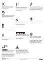 3
3
-
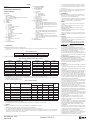 4
4
-
 5
5
-
 6
6
-
 7
7
-
 8
8
-
 9
9
-
 10
10
-
 11
11
-
 12
12
-
 13
13
-
 14
14
-
 15
15
-
 16
16
-
 17
17
-
 18
18
-
 19
19
-
 20
20
-
 21
21
-
 22
22
-
 23
23
-
 24
24
-
 25
25
-
 26
26
-
 27
27
-
 28
28
-
 29
29
-
 30
30
-
 31
31
-
 32
32
-
 33
33
-
 34
34
-
 35
35
-
 36
36
-
 37
37
-
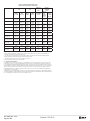 38
38
-
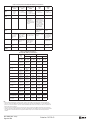 39
39
-
 40
40
-
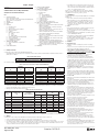 41
41
-
 42
42
-
 43
43
-
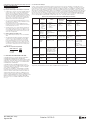 44
44
-
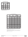 45
45
-
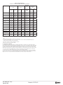 46
46
-
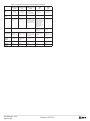 47
47
-
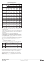 48
48
-
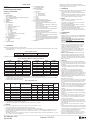 49
49
-
 50
50
-
 51
51
-
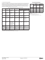 52
52
-
 53
53
-
 54
54
-
 55
55
-
 56
56
-
 57
57
-
 58
58
-
 59
59
-
 60
60
-
 61
61
-
 62
62
-
 63
63
-
 64
64
-
 65
65
-
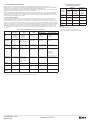 66
66
-
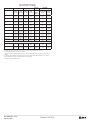 67
67
-
 68
68
-
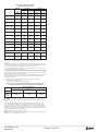 69
69
-
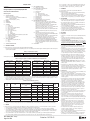 70
70
-
 71
71
-
 72
72
-
 73
73
-
 74
74
-
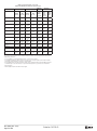 75
75
-
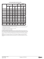 76
76
-
 77
77
-
 78
78
-
 79
79
-
 80
80
-
 81
81
-
 82
82
-
 83
83
-
 84
84
-
 85
85
-
 86
86
-
 87
87
-
 88
88
-
 89
89
-
 90
90
-
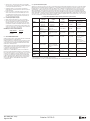 91
91
-
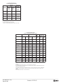 92
92
-
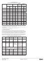 93
93
-
 94
94
-
 95
95
-
 96
96
-
 97
97
-
 98
98
-
 99
99
-
 100
100
-
 101
101
-
 102
102
-
 103
103
-
 104
104
-
 105
105
-
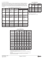 106
106
-
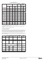 107
107
-
 108
108
-
 109
109
-
 110
110
-
 111
111
-
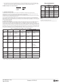 112
112
-
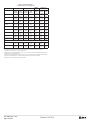 113
113
-
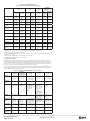 114
114
-
 115
115
-
 116
116
-
 117
117
-
 118
118
-
 119
119
-
 120
120
-
 121
121
-
 122
122
-
 123
123
-
 124
124
-
 125
125
-
 126
126
-
 127
127
-
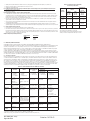 128
128
-
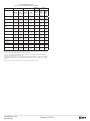 129
129
-
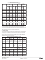 130
130
-
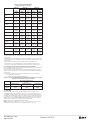 131
131
-
 132
132
-
 133
133
-
 134
134
-
 135
135
-
 136
136
-
 137
137
-
 138
138
-
 139
139
-
 140
140
-
 141
141
-
 142
142
-
 143
143
-
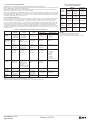 144
144
-
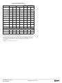 145
145
-
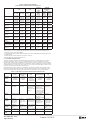 146
146
-
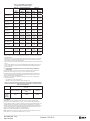 147
147
-
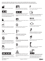 148
148
-
 149
149
-
 150
150
-
 151
151
-
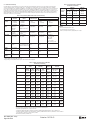 152
152
-
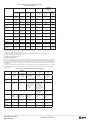 153
153
-
 154
154
-
 155
155
-
 156
156
-
 157
157
-
 158
158
-
 159
159
-
 160
160
-
 161
161
-
 162
162
-
 163
163
-
 164
164
-
 165
165
-
 166
166
-
 167
167
-
 168
168
-
 169
169
-
 170
170
-
 171
171
-
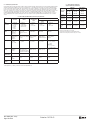 172
172
-
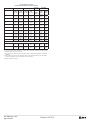 173
173
-
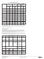 174
174
-
 175
175
-
 176
176
-
 177
177
-
 178
178
-
 179
179
-
 180
180
-
 181
181
-
 182
182
-
 183
183
-
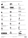 184
184
-
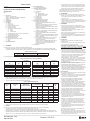 185
185
-
 186
186
-
 187
187
-
 188
188
-
 189
189
-
 190
190
-
 191
191
-
 192
192
-
 193
193
-
 194
194
-
 195
195
-
 196
196
-
 197
197
-
 198
198
-
 199
199
-
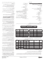 200
200
-
 201
201
-
 202
202
-
 203
203
-
 204
204
-
 205
205
-
 206
206


în alte limbi
- slovenčina: Abbott XIENCE Sierra Používateľská príručka
Alte documente
-
 Merit Medical Arcadia Steerable Balloon and Straight Balloon Catheter Instrucțiuni de utilizare
-
 Merit Medical RadialFlo Instructions For Use Manual
-
Bard Magic3 Go Instructions For Use Manual
-
 Johnson & Johnson Biosense Webster ThermoCool SmartTouch Instructions For Use Manual
-
Vents Z,Vitro,Z star,Vitro star Manual de utilizare
-
Laserliner DampFinder Compact Manualul proprietarului
-
Covidien Shiley 137880 Manual de utilizare
-
Arrow Peripherally Inserted Central Instrucțiuni de utilizare
-
-
 argon audio BioPince Instructions Manual


